Implantology
Dental Restoration
Digital Dentistry
Dental Labrotary
Socket Lift and Implant Placement in the right maxilla.

HAPPYTOGETHER
Views : 3,940/ Jan, 19, 2023
Views : 3,940/ Jan, 19, 2023
<GCacg> A 56-year-old male patient complained of pain in the right upper and lower jaws. And he wanted the upper first molar to be pulled out first.
 ▲Initial intraoral view
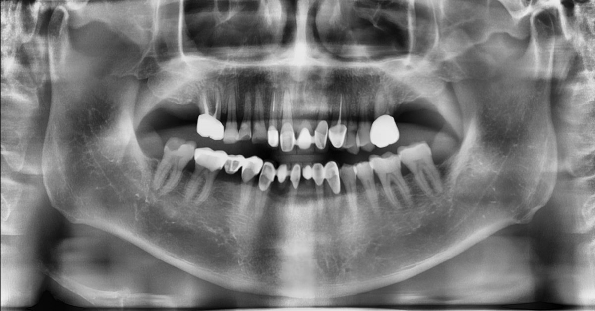 ▲Panoramic radiograph
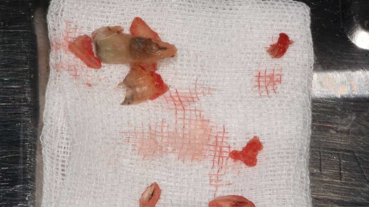 ▲The troubled tooth was removed in the first place.
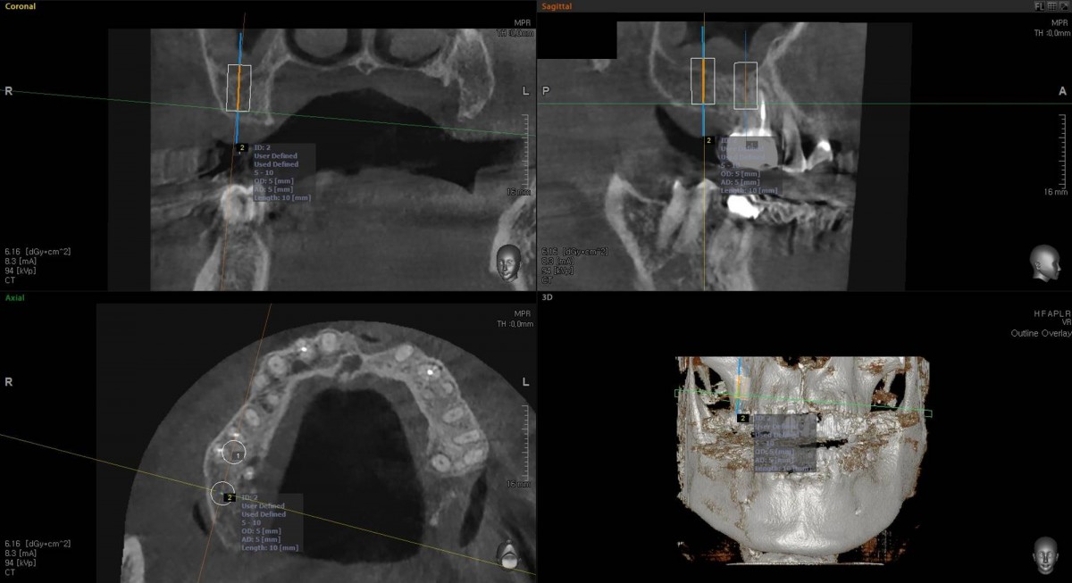 ▲Surgery simulation on the OBCT image before implant placement
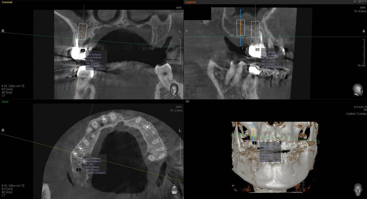 ▲Surgery simulation on the OBCT image before implant placement
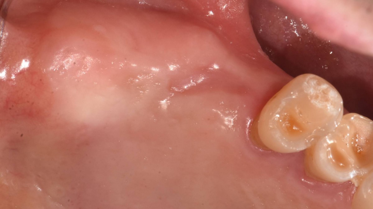 ▲On the day of implant placement, intra-oral view after 2months of 1st molar extraction.
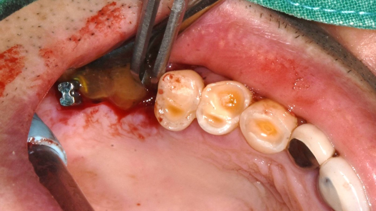 ▲CGF was applied after osteotomy using sinus reamers.
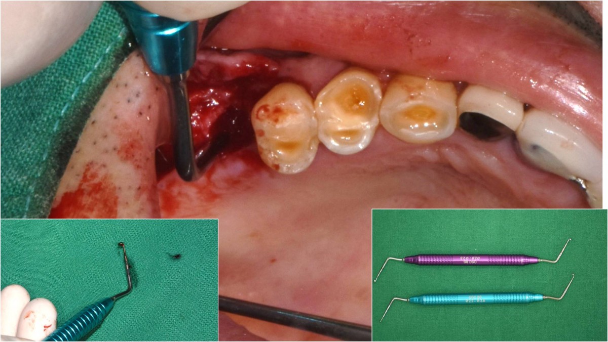 ▲The Membrane-Lifter was applied to lift the sinus membrane.
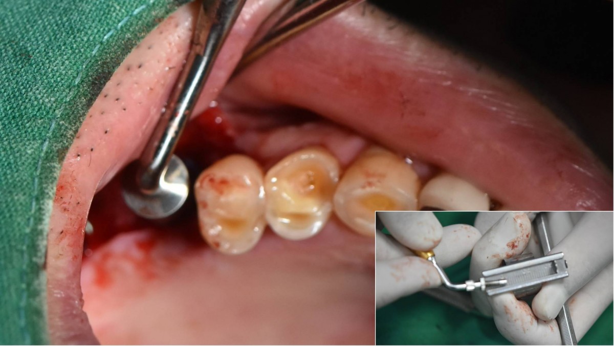 ▲Choice of graft material was applied into the osteotomy site then was pushed up using a Bone-Packer and Sinus-Lifter.
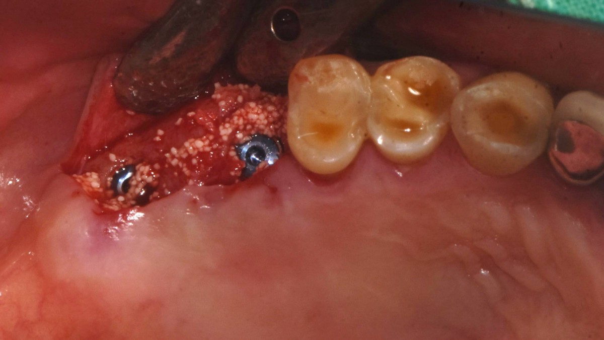 ▲ 2 implants were placed in position with Arum-Dentistry NB-1 Ø5.0/L10mm. Initial stability was 50Ncm in the 1st molar area and 40Ncm in the 2nd molar area.
 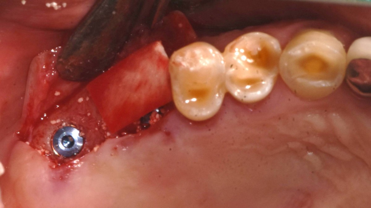 ▲The grafted site was covered with a collagen membrane
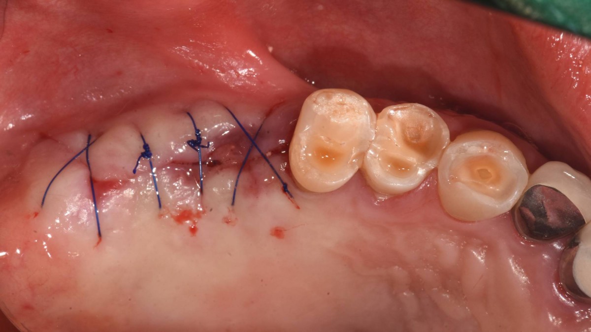 ▲The flap was closed.
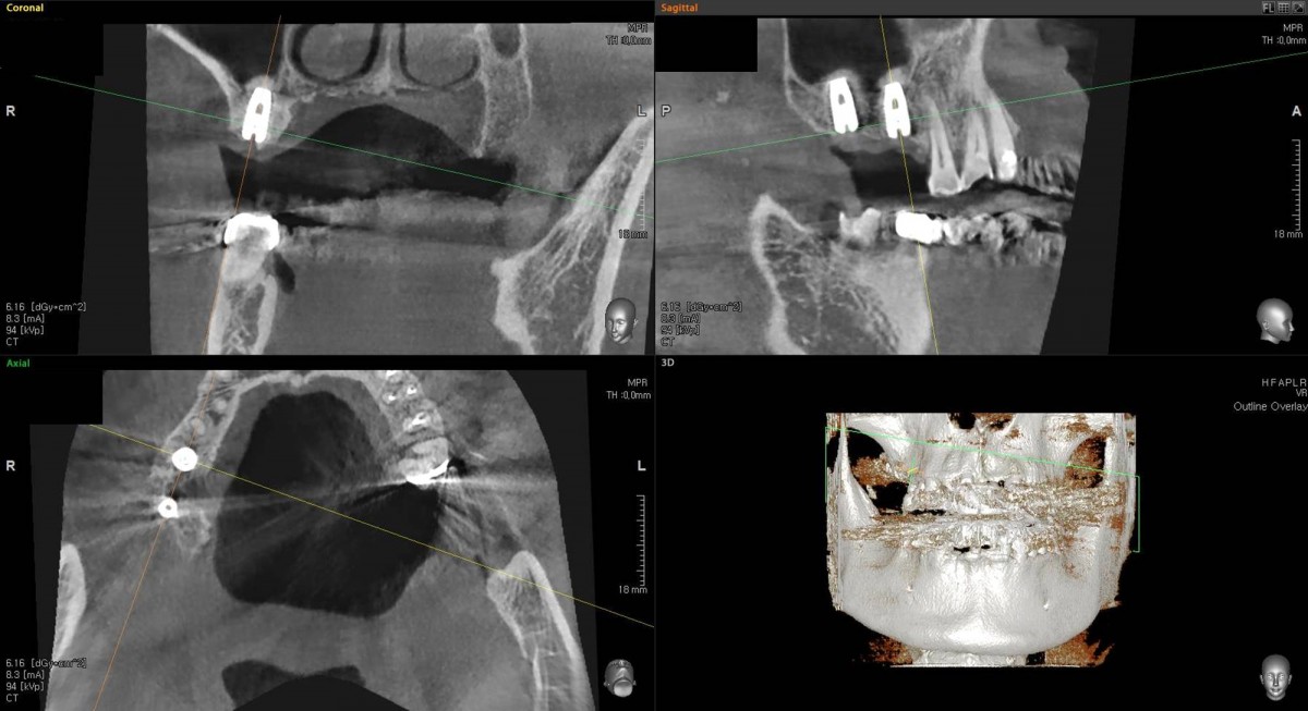 ▲CBCT scan image focused on 1st molar zone
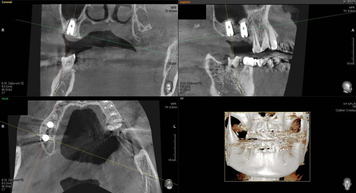 ▲CBCT scan image focused on 2nd molar zone,
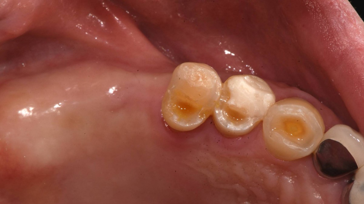 ▲4.5months post-op. Intra-oral photo on the day of implant uncovery(2nd surgery).
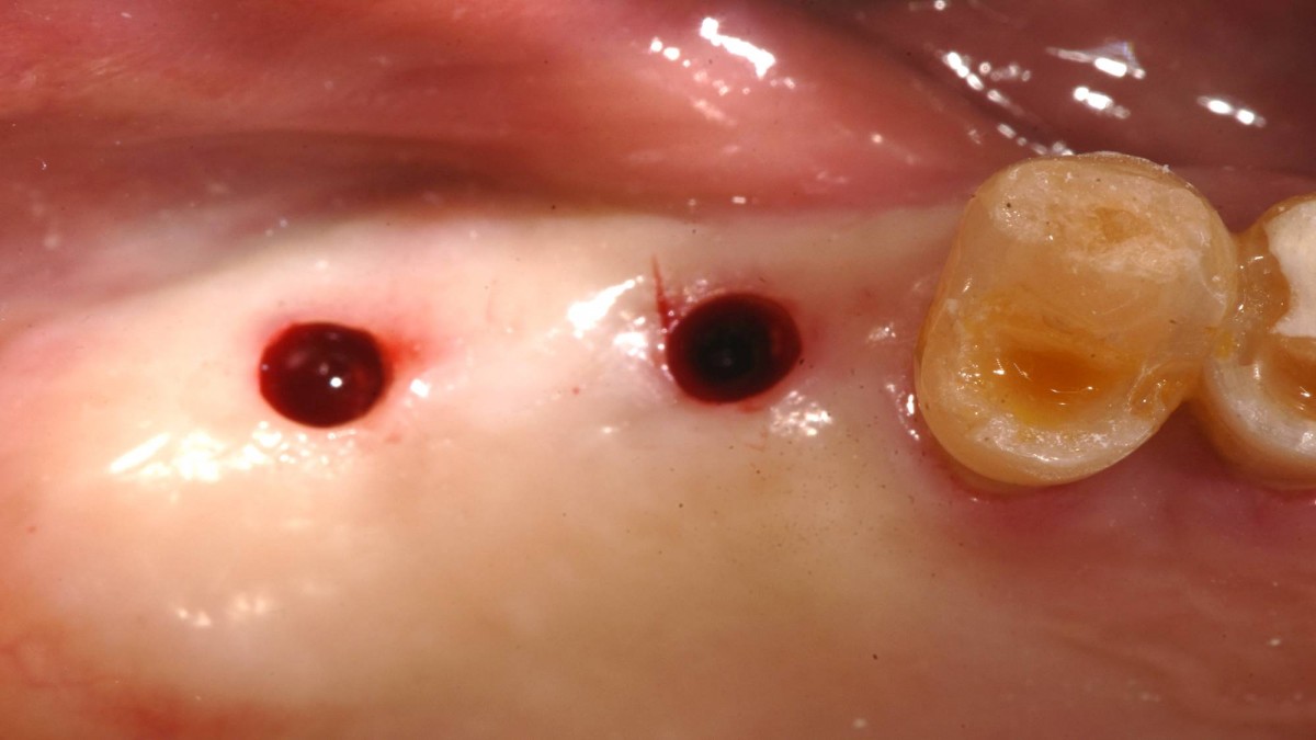 ▲Tissue punching was done
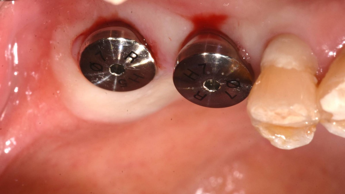 ▲Healing abutments were engaged to the implant
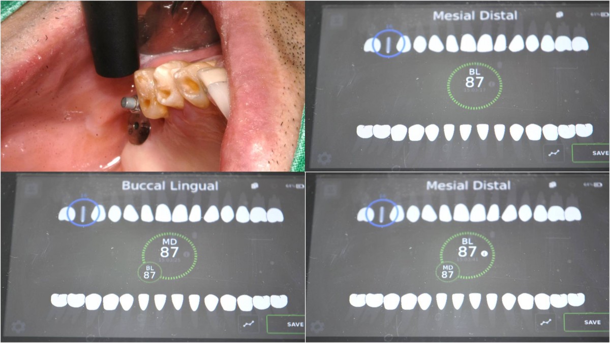 ▲ISQ value reading
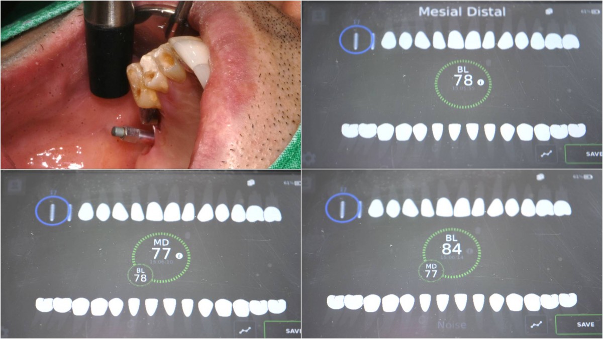 ▲ISQ value reading
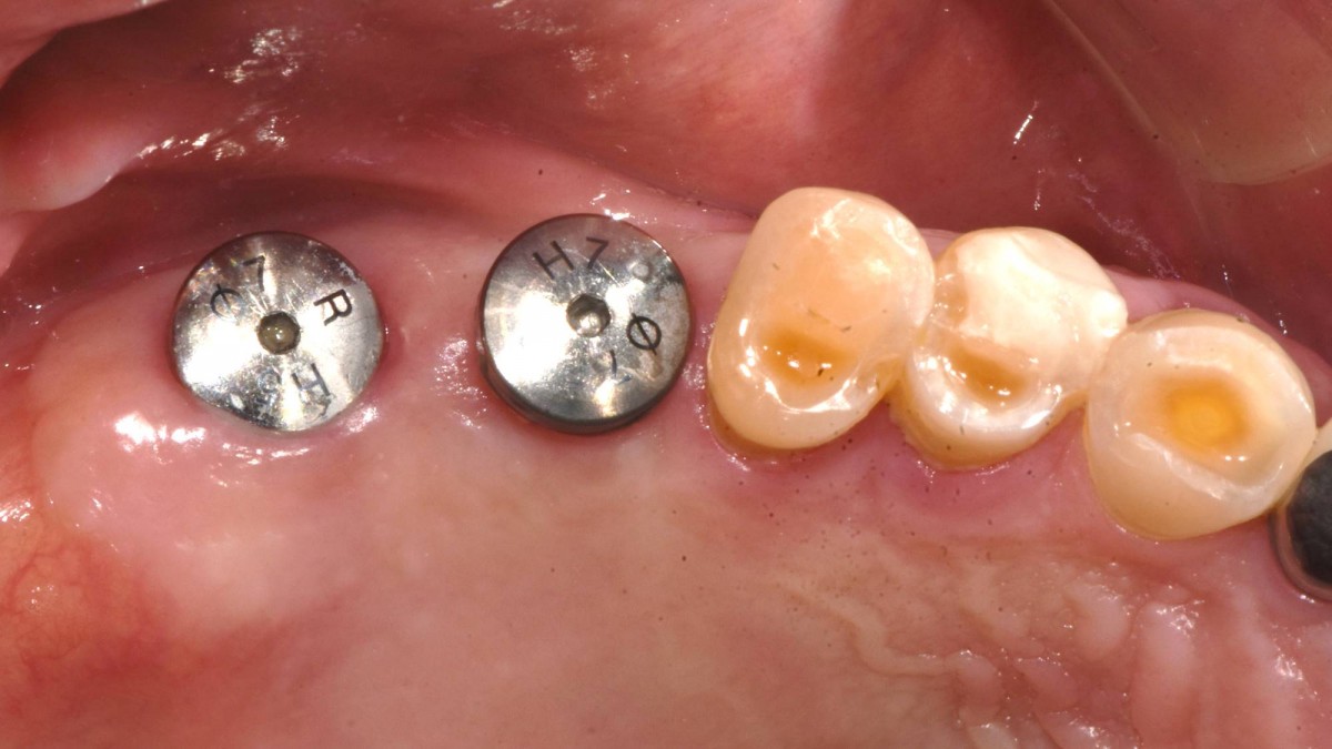 ▲5months post-op. The healing abutments would be switched with scan abutments for intra-oral scanning.
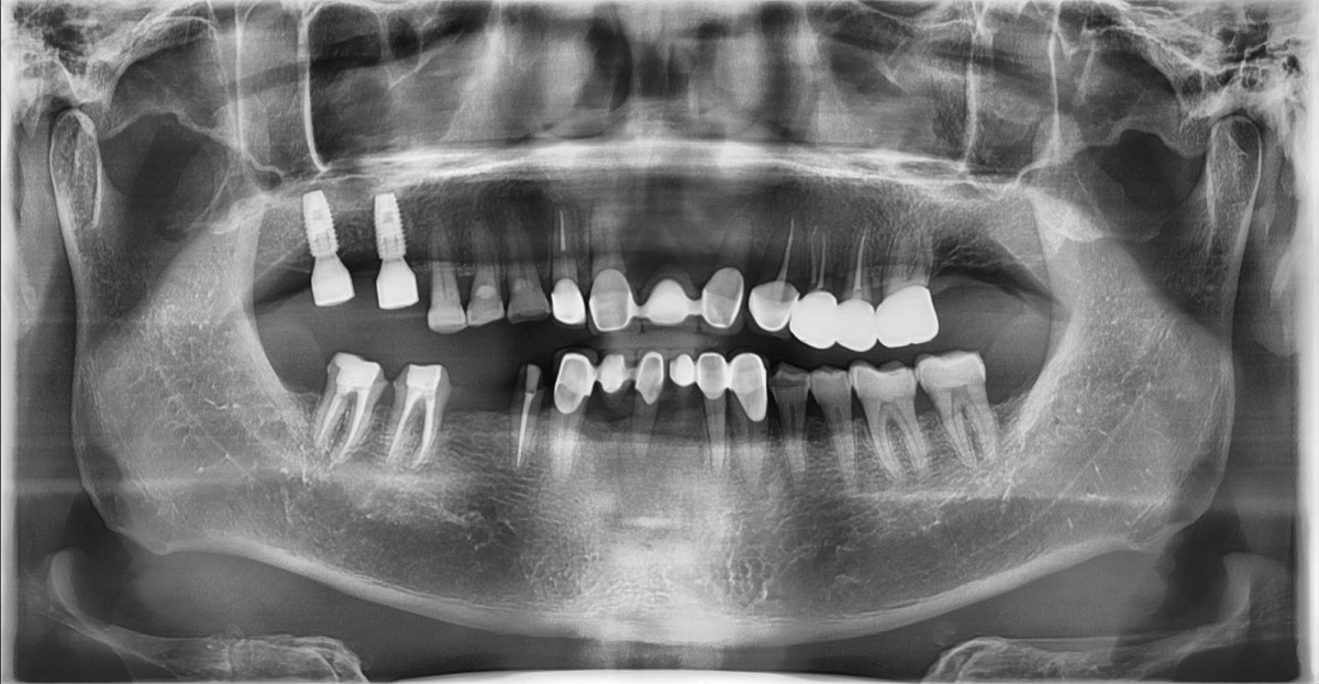 ▲scan abutments were connected and a panoramic radiograph was taken to make sure of the connection.
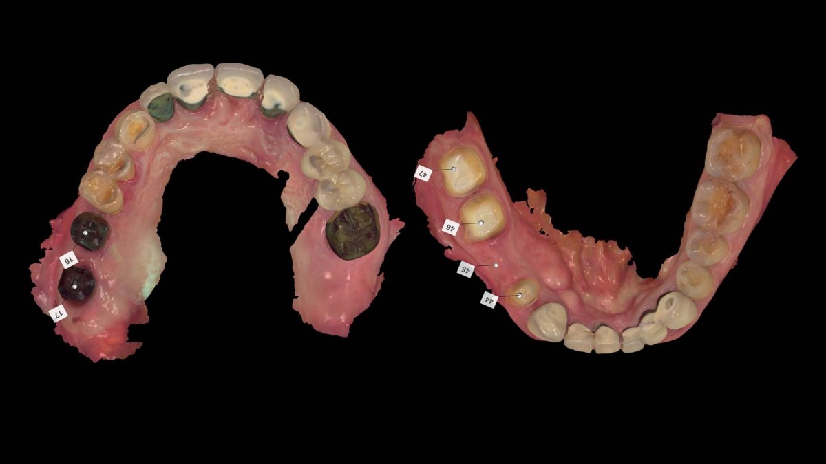 ▲Intraoral scanning for digital impression
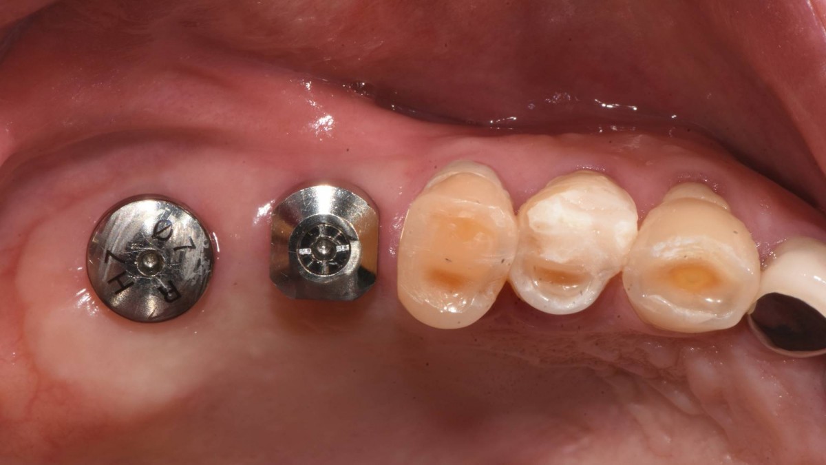 ▲The patient complained of some sharpness and it hurt his tongue due to a scan abutment in the 2nd molar zone. That was replaced with a healing abutment after intra-oral scanning.
 ▲Customized abutments, orientation jig, and prosthesis
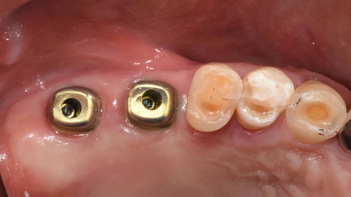 ▲Custom abutments were connected to the fixtures.
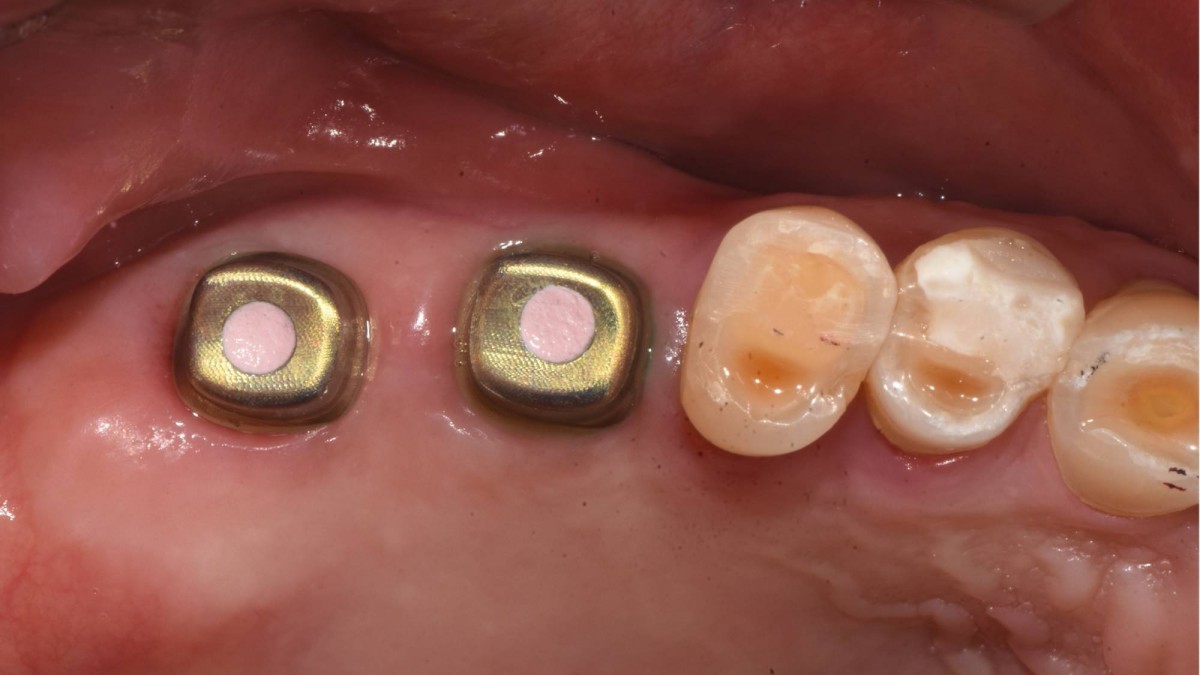 ▲The screw holes were filled with temporary filling material
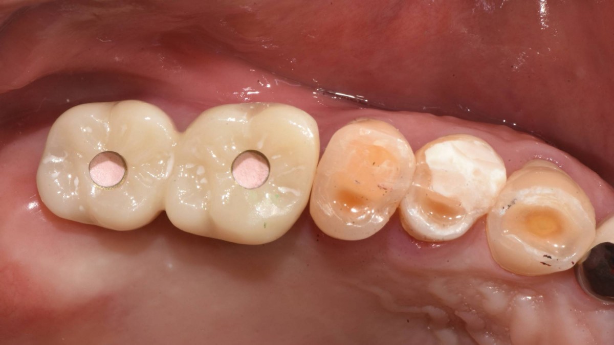 ▲Crown seating trial
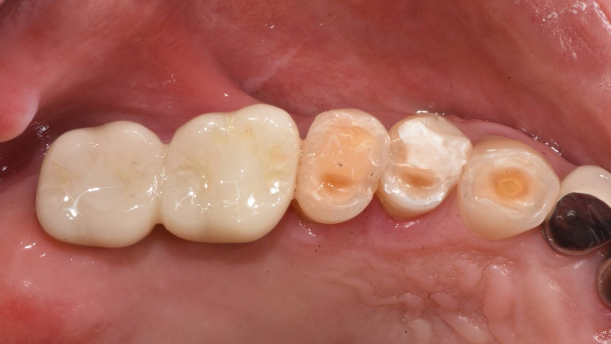 ▲The crown was cemented permanently and the access hole was filled with composite resin.
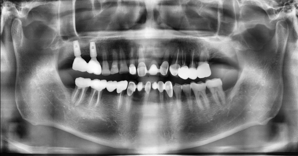 ▲Panoramic radiograph was taken after restoration delivery
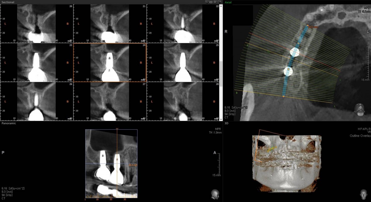 ▲CBCT after restoration delivery
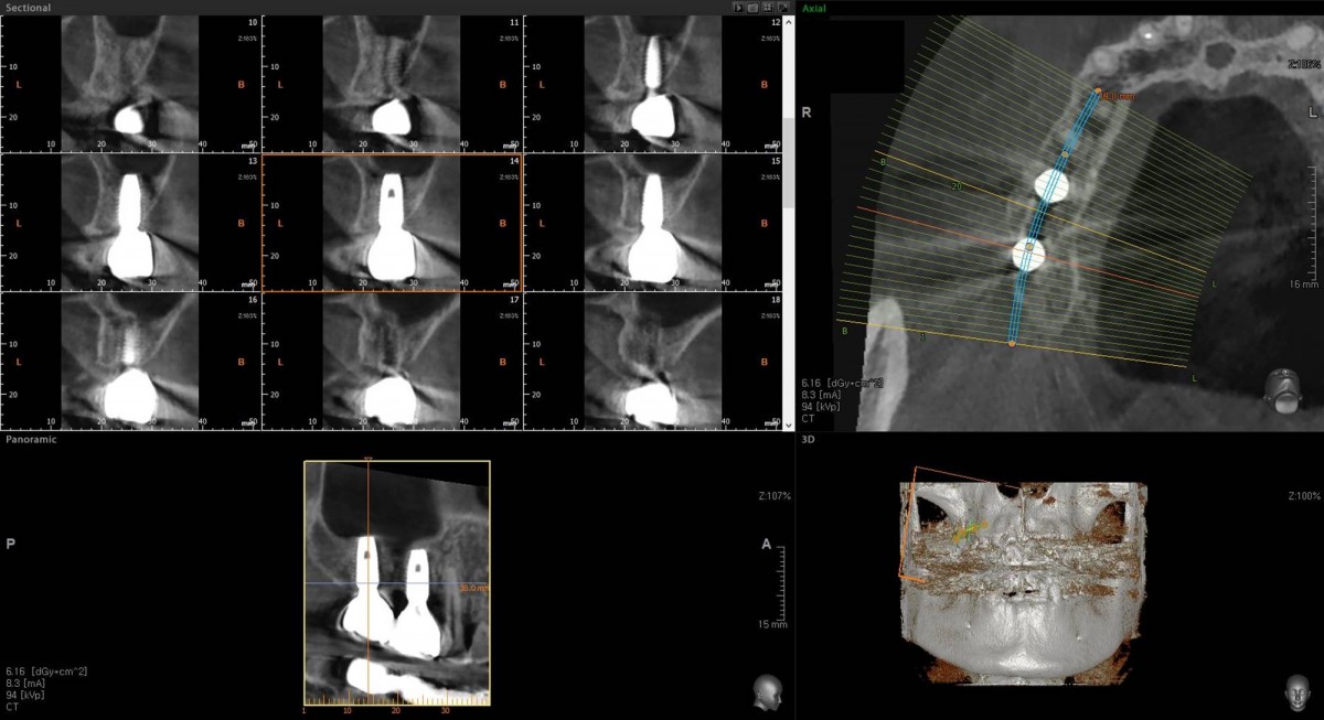 ▲CBCT after restoration delivery
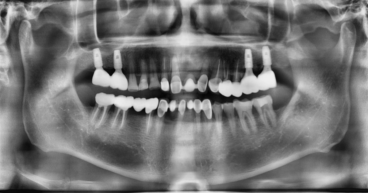 ▲panoramic radiograph. almost 1 year later.
0
- PrevIn the anterior maxilla, implant-supported fixed partial denture.Jan, 19, 2023
- NextImmediate implant placement in the left molar of the maxilla. Jan, 19, 2023
There are no registered comment.


