

Implantology
Dental Restoration
Digital Dentistry
Dental Labrotary
3 implants in the Mandible, Arum implant.

Views : 4,790/ Dec, 06, 2022
A 62-year-old female had heart
disease and angiostenosis with the daily taking of a thrombolytic agent. And
even harder was she got a pain intolerance. She showed multiple missing areas
in the molar region and almost hopeless remaining teeth with mobility.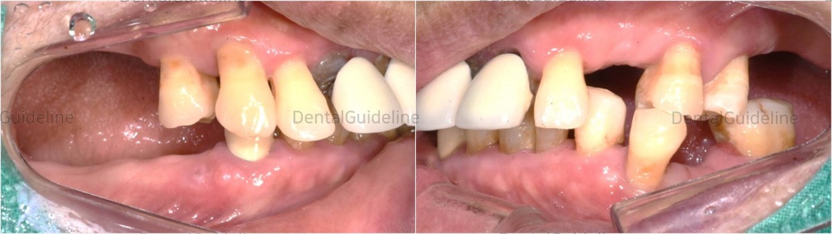
Intra-oral photo before implant surgery.

Pre-op
panoramic radiograph.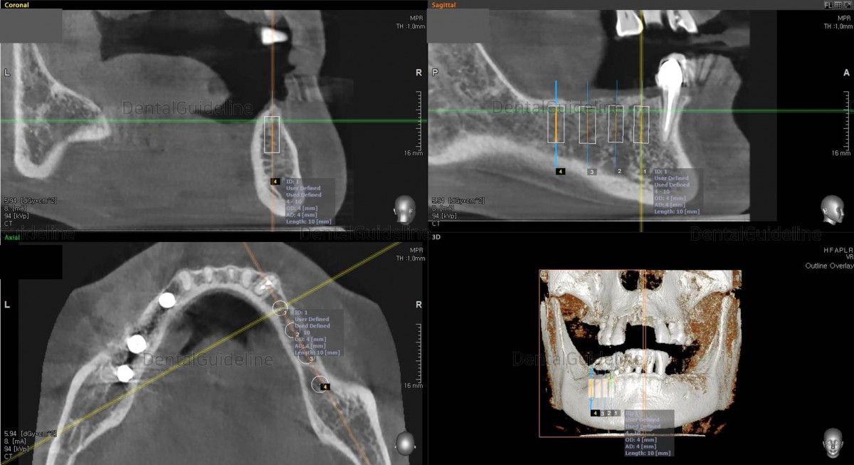
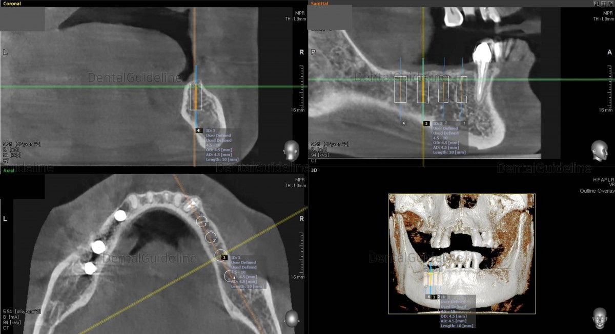
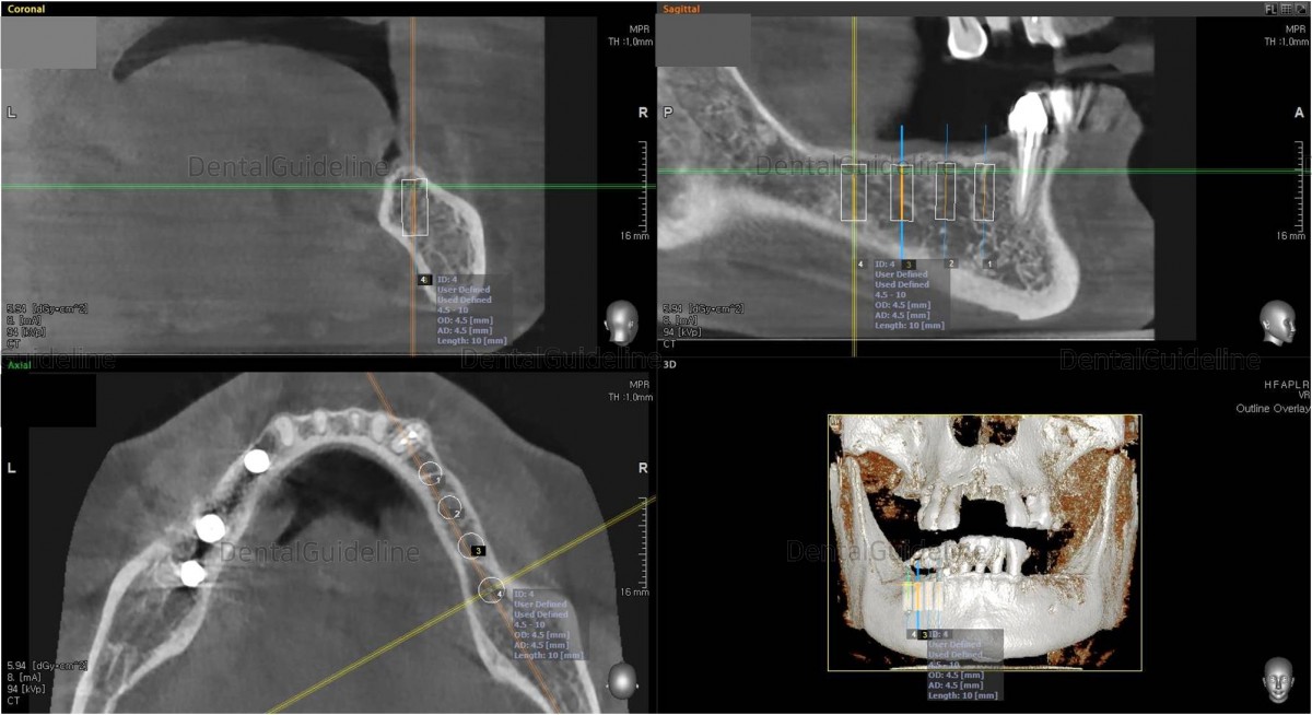
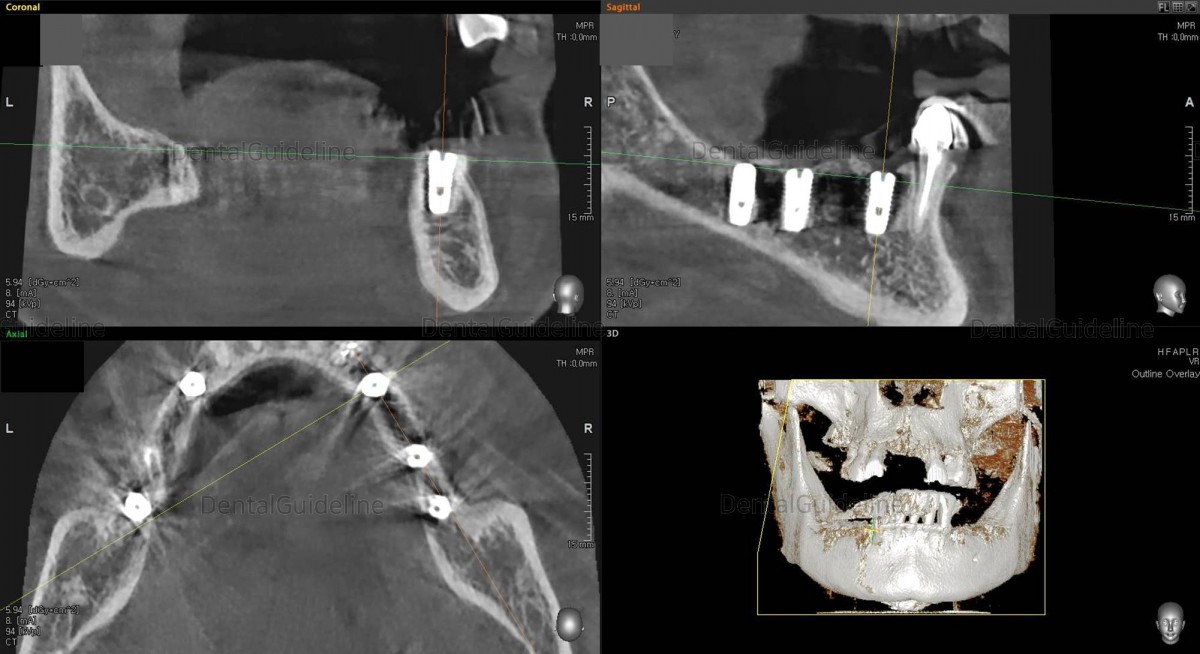
Simple
simulation of implant placement on the CBCT scan image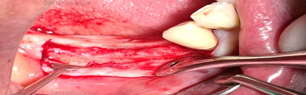
The flap was opened to expose the
surgical site.
Serial drilling.
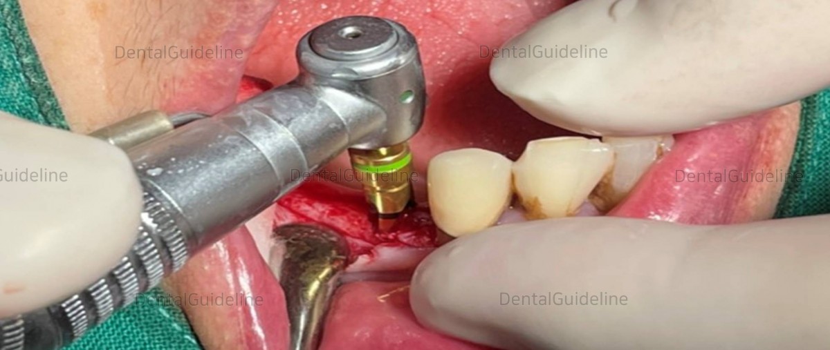
Final installation with favorable initial
stability.

3
implants were placed on the lower-left area and additive GBR was tried., 
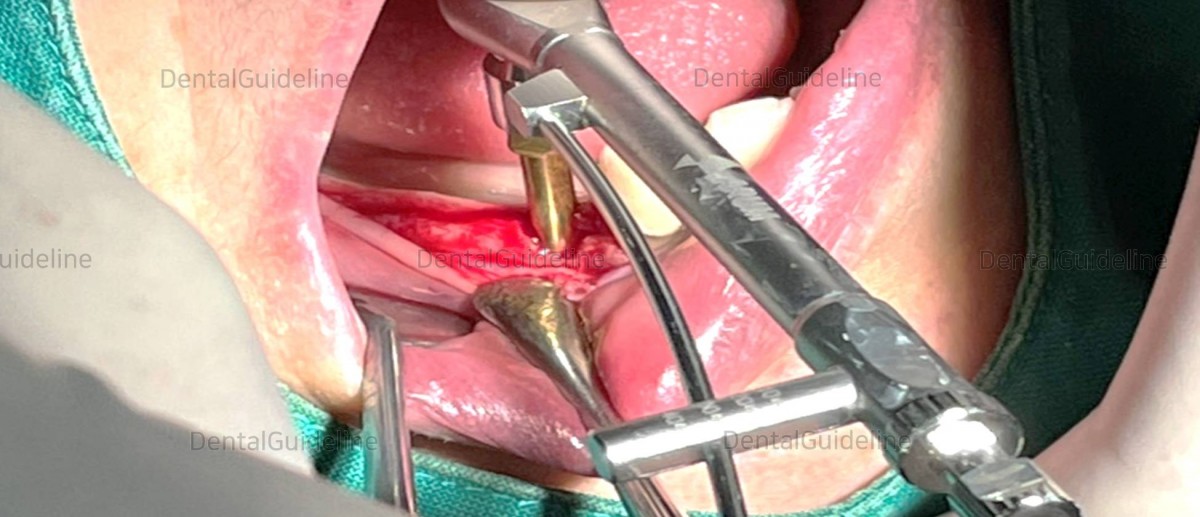
A collagen membrane was covered
over the surgical site.

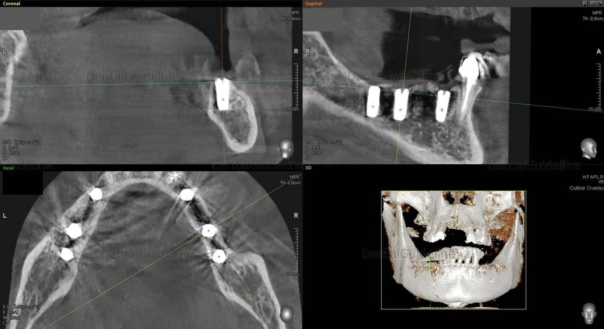
Post-op panoramic radiograph.
CBCT
scan. Arum NB-1 Ø4.0/L10
(30Ncm) at the 1st premolar zone.
CBCT scan Arum NB-1 Ø4.5./ L10
(20Ncm) at
the 1st molar zone.
CBCT scan Arum NB-1 Ø4.5/ L10
(20Ncm) at
the 2nd molar zone.Arm Dentistry NB-1 Ø4.5/ L10
(20Ncm) at
the 2nd molar zone.Arum Dentistry NB-1 Ø4.5/ L10
(20Ncm) at
the 2nd molar zone.
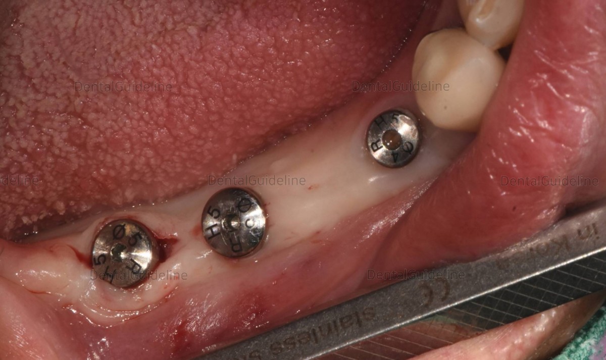
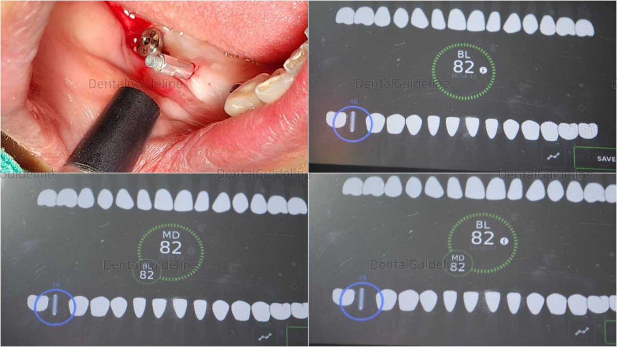
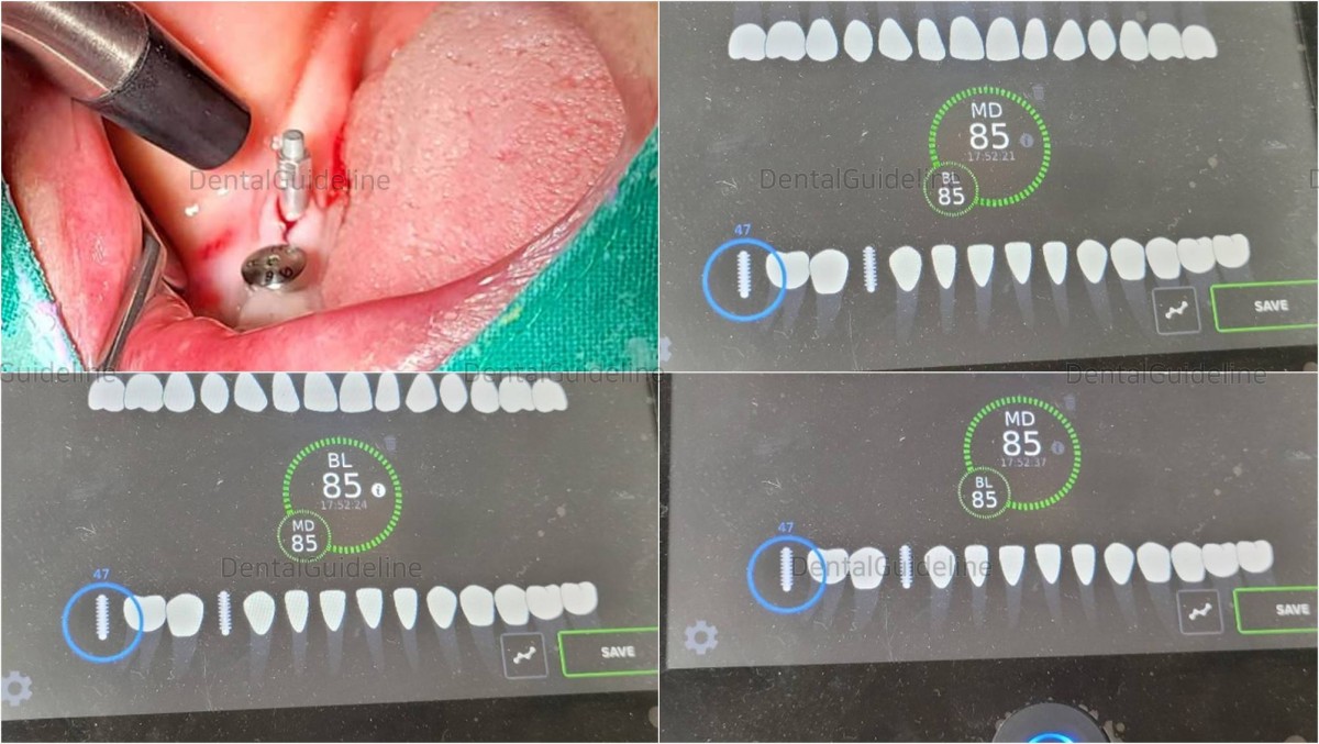
On the day of implant uncovery and ISQ reading, a small-sized healing abutment was engaged first to secure the gingival tissue by expanding gradually.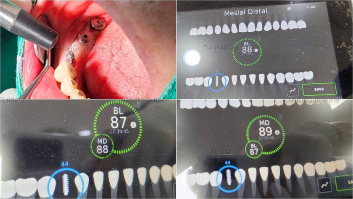
ISQ reading in the 1st premolar zone.
ISQ reading in the 1st molar zone
ISQ reading in the 2nd molar zone.
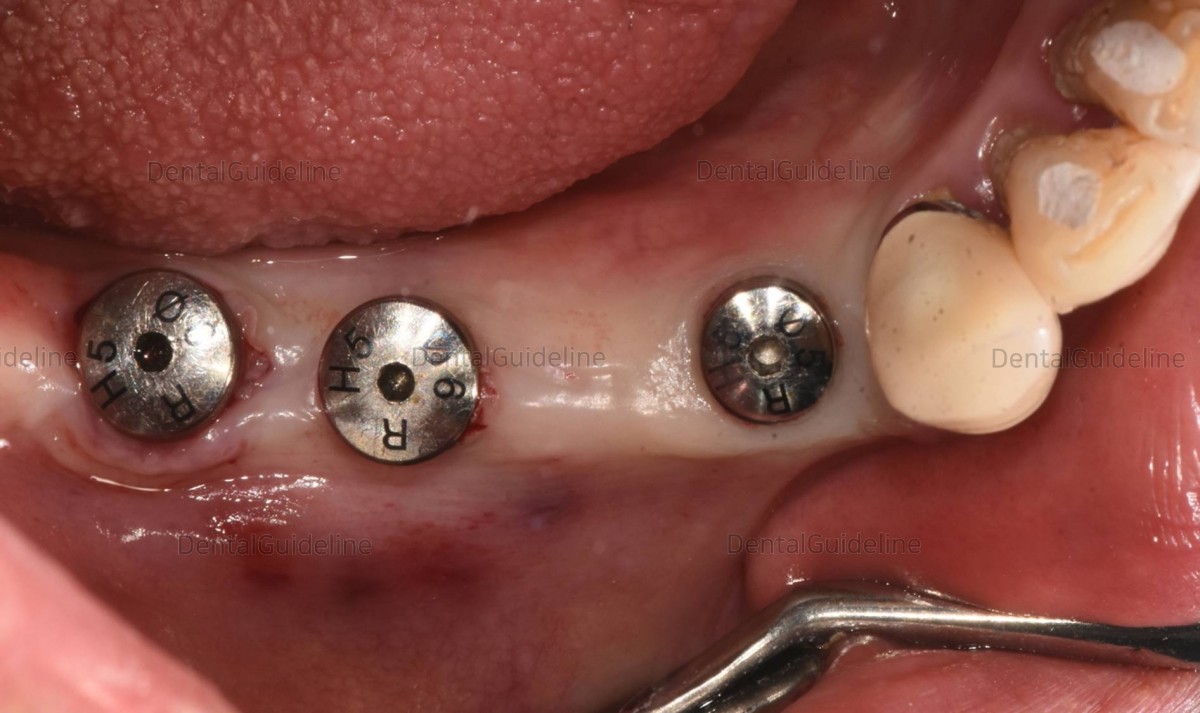
Final-sized healing abutments were engaged to the fixture after gradual
expansion using the serial size of the healing abutment on the day of the implant 2nd surgery and ISQ
reading.
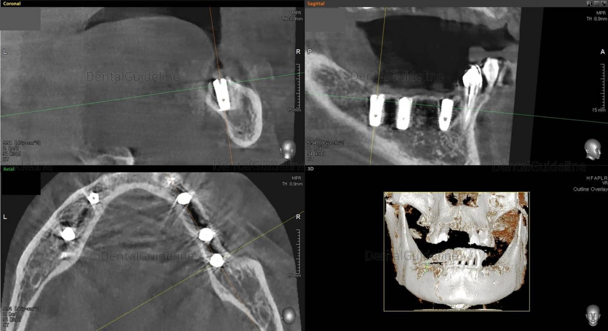
4 days after the implant second surgery.
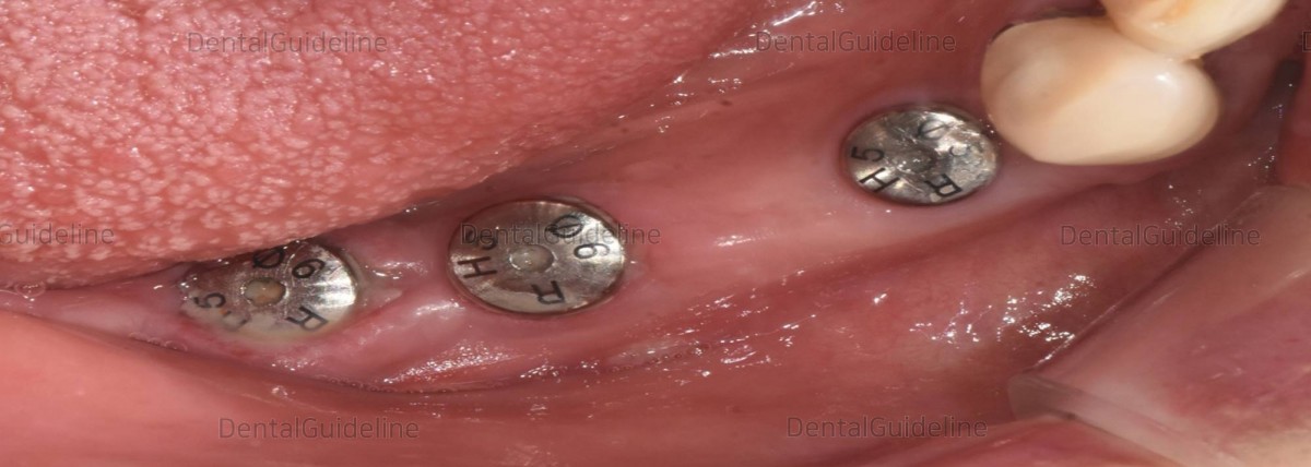
6 weeks
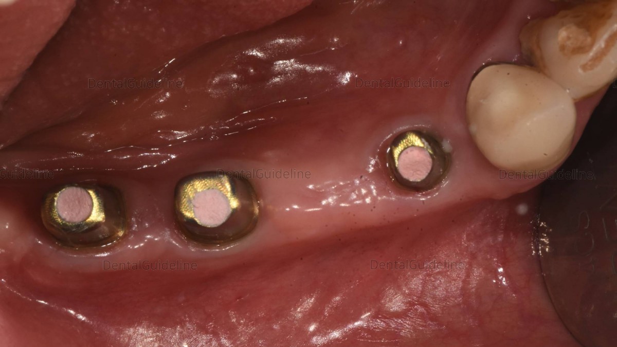
after the implant 2nd surgery, healing abutments were replaced by scan abutments for intra-oral scanning.
A panoramic radiograph was taken to check
the secure connection of scan abutments.
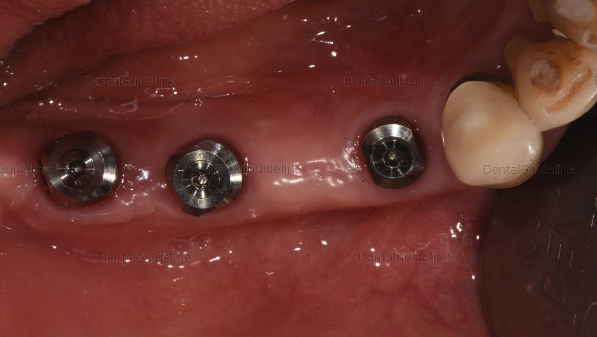
Intra-oral scanned image for digital impression.
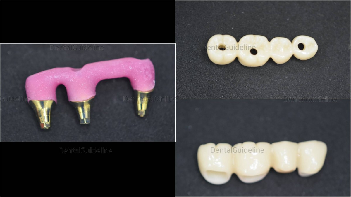
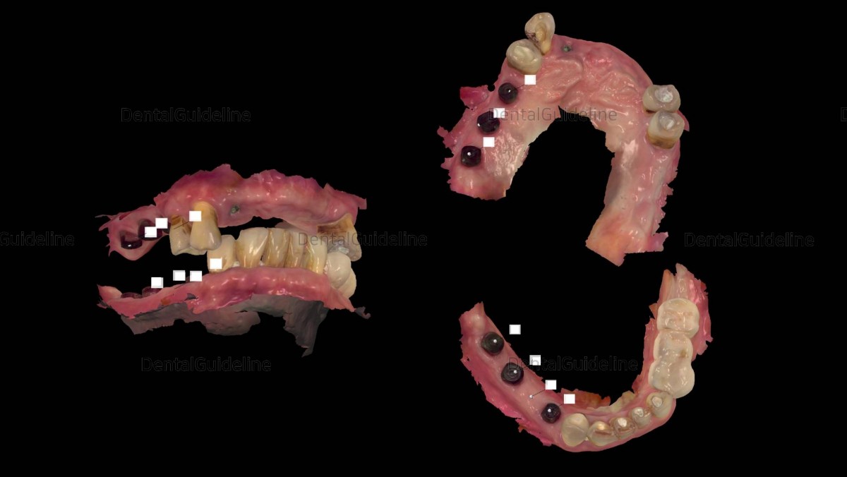
Custom abutments, orientation jig, and
restoration.
Abutments were connected to fixtures and
screw holes were filled with the temporary filling material for retrievability.
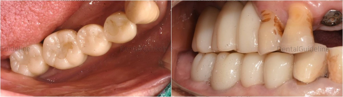
Intra-oral photos were taken after
2 months of restoration.

A panoramic radiograph was taken after 2 months of loading

A panoramic radiograph was taken after 13 months of loading
- PrevImplant in the upper and lower second molarDec, 06, 2022
- NextImmediate placement of 3 implants, Scanning, ISQ measurement. Arum implant system. Dec, 06, 2022
There are no registered comment.