Endodontics
Oral Surgery
Apical Surgery (Apicoectomy) and bone graft

HAPPYTOGETHER
Views : 1,502/ Nov, 06, 2022
Views : 1,502/ Nov, 06, 2022
Apical lesion at the premolar. <Ghsr>
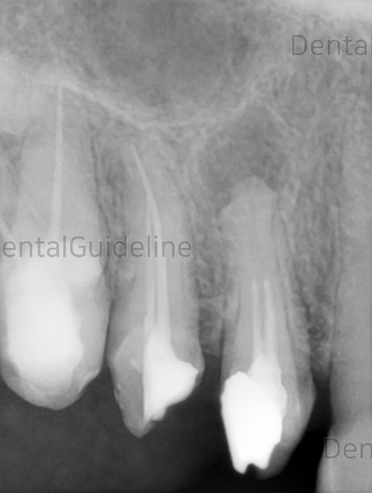
The periapical radiograph shows an apical lesion with incomplete root canal treatment at the root apex of the first premolar.
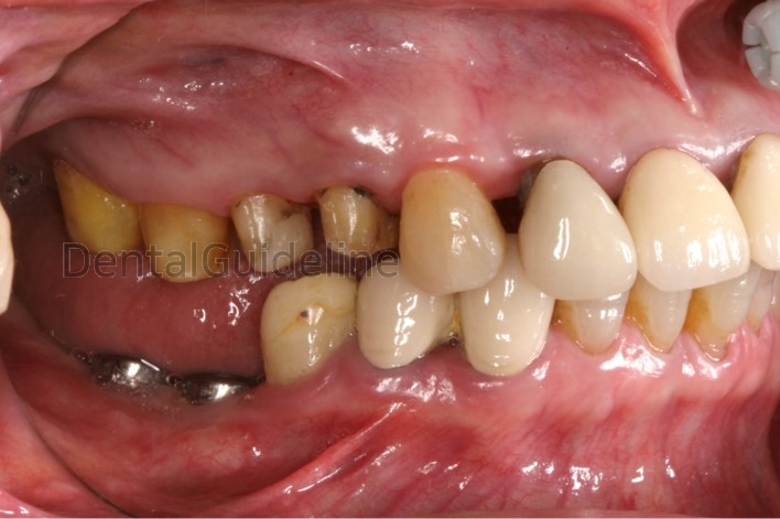
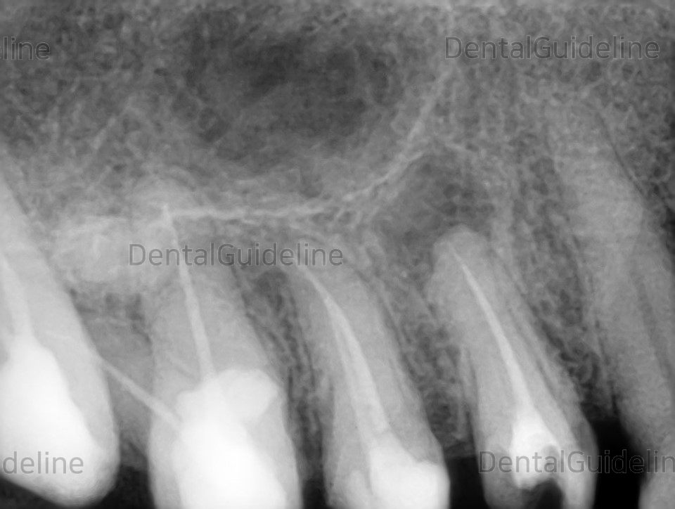
Root canal re-treatment was done first.
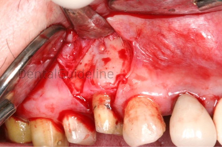
Exposed lesion
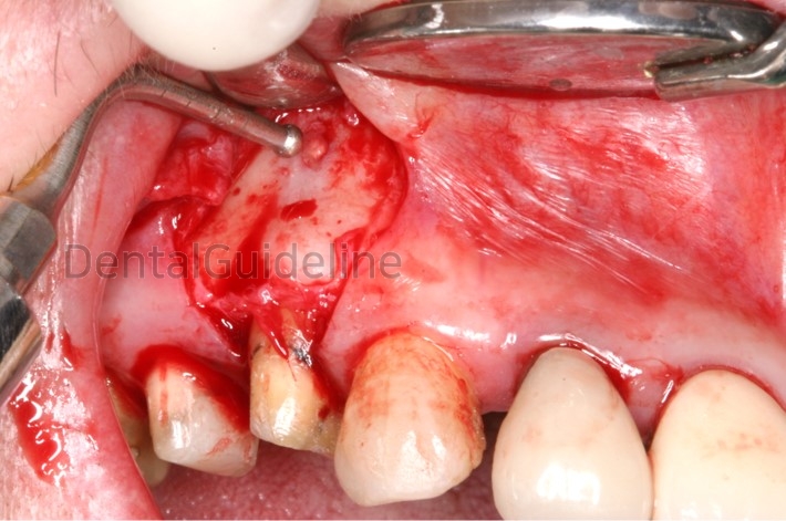
Osteotomy and removal of granulation tissue with SURGYBONE®
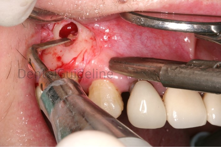
Cut the infected root apex.
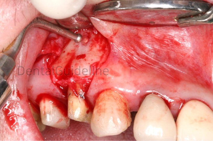
Trimming of the shart area. The retrograde filling was not tried.
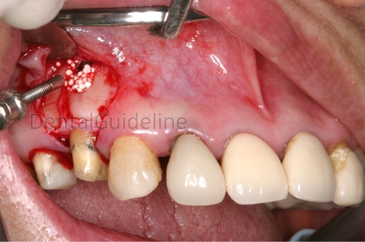
The lesion was filled with synthetic bone (beta-TCP).
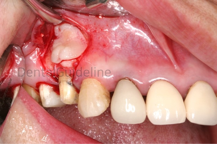
Instead of collagen membrane, CGF(Concentrated Growth Factors) was used to cover the grafted site.
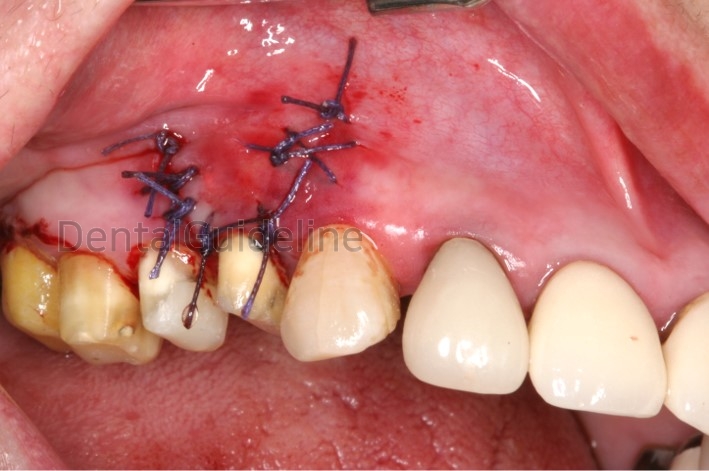
Suture
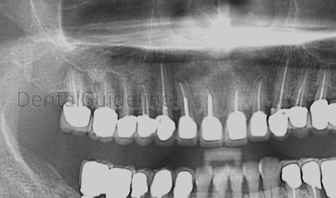
Post=op radiograph.

The periapical radiograph shows an apical lesion with incomplete root canal treatment at the root apex of the first premolar.


Root canal re-treatment was done first.

Exposed lesion

Osteotomy and removal of granulation tissue with SURGYBONE®

Cut the infected root apex.

Trimming of the shart area. The retrograde filling was not tried.

The lesion was filled with synthetic bone (beta-TCP).

Instead of collagen membrane, CGF(Concentrated Growth Factors) was used to cover the grafted site.

Suture

Post=op radiograph.
0
- PrevFlapless Surgery and Attached Gingiva Control, Tissue Punch.Nov, 06, 2022
- NextUseful tips for making maxillary denture. Reproduction of anatomical shape Nov, 06, 2022
There are no registered comment.


