Implantology
Dental Restoration
Dental Labrotary
Immediate placement, GBR, Arum NB-1

HAPPYTOGETHER
Views : 2,965/ Dec, 12, 2022
Views : 2,965/ Dec, 12, 2022
<GCkhk>
A 56-year-old male patient had
repeated painful experiences during chewing.
The tooth that showed crack-tooth syndrome and
furcation-involved perio problem was scheduled to be extracted and immediate
placement of an implant.
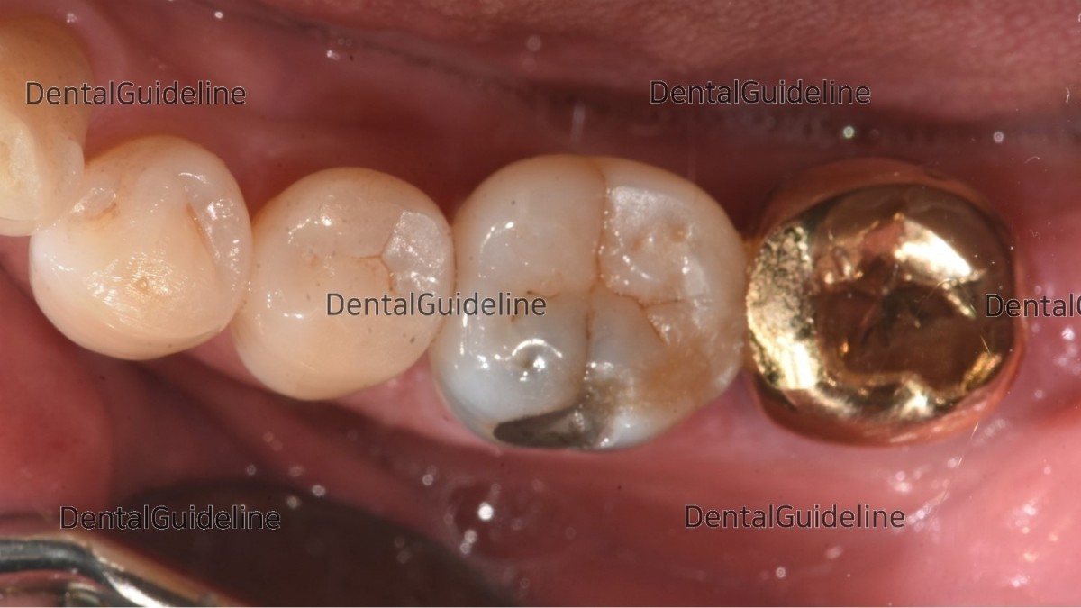
Lower left 1st molar had a distal crack, secondary caries around old filling material, and caries with furcation involvement.
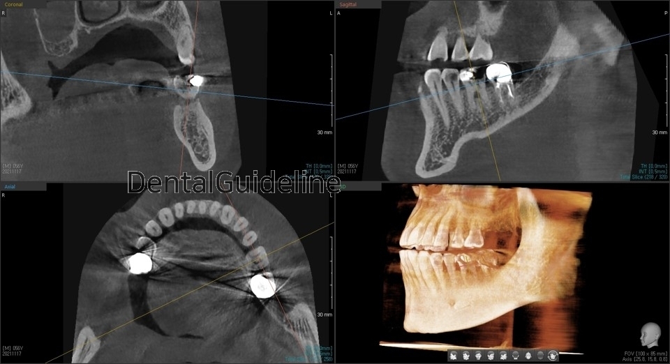
CBCT scanning image focused on the 1st molar
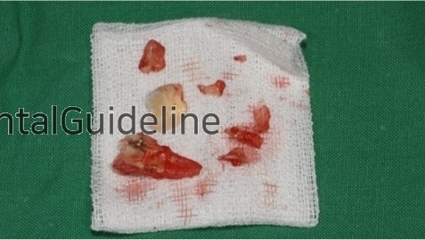
extraction
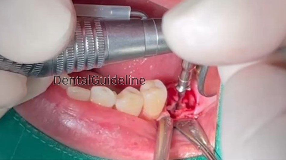
Serial drilling
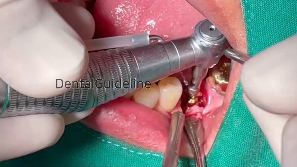
Serial drilling
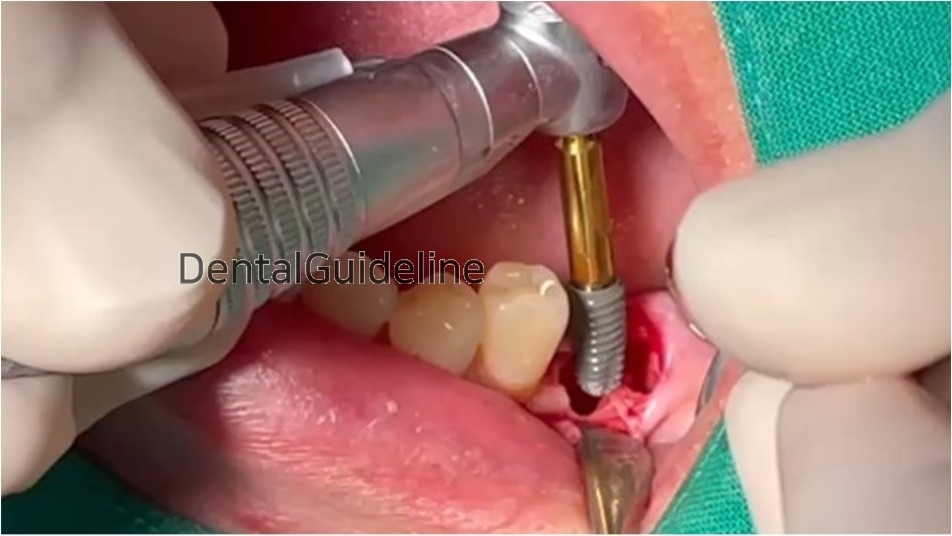
The implant was ready to be placed into the osteotomy site.
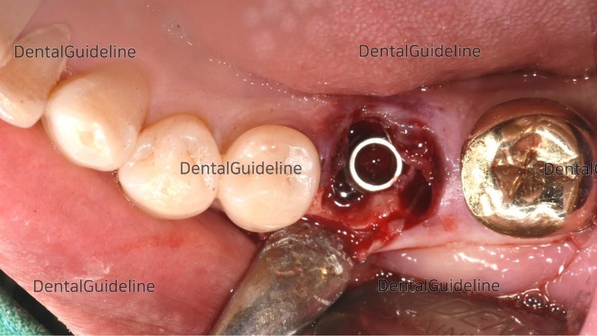
The implant was placed in position.
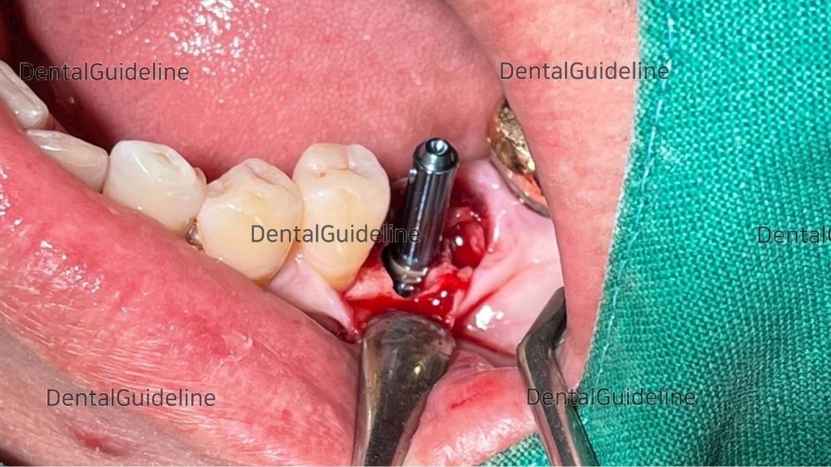
The Direction-Pin was engaged in the implant to check the 3-dimensional position.
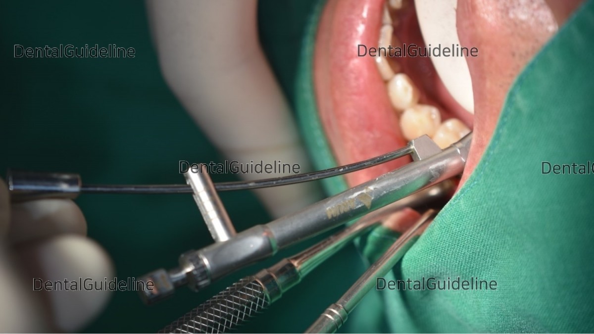
Confirm the final insertion. The initial stability was good enough(40Ncm).
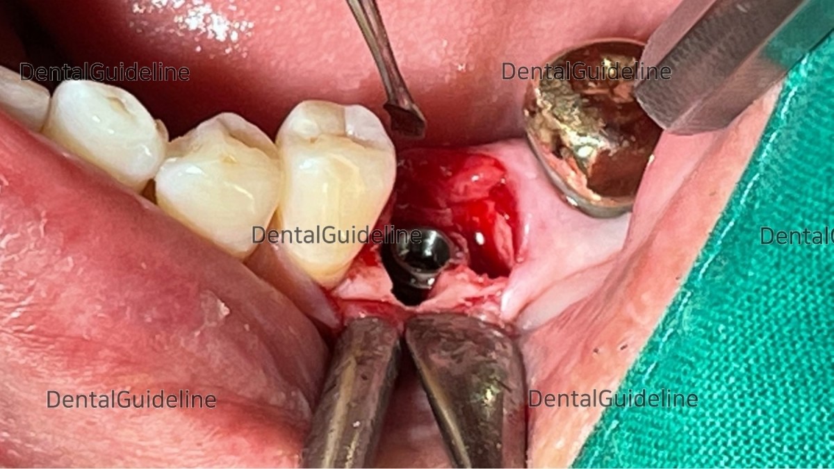
The implant was placed in the final position. Arum NB-1 Ø5.0/L10mm.
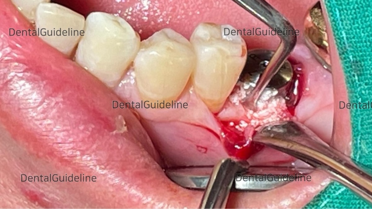
Grafting procedure with synthetic bone (beta-TCP).
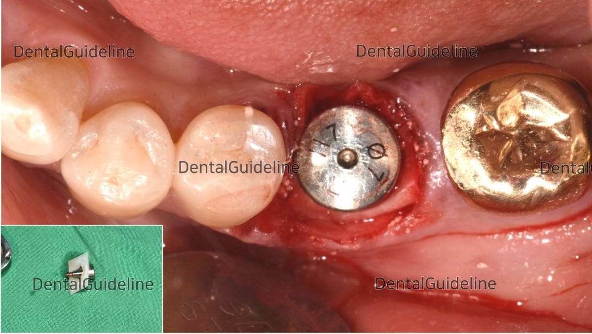
The collagen membrane-healing abutment assembly was connected to the fixture.
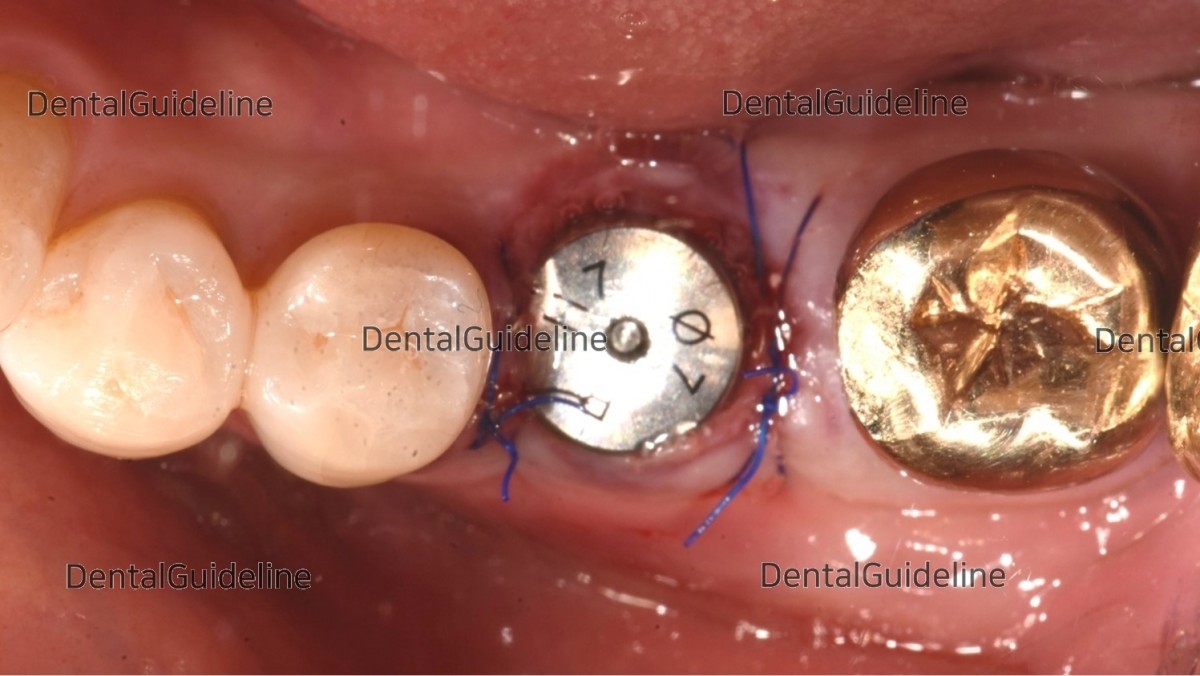
suture (blue nylon).
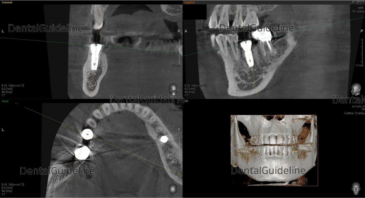
CBCT scan image after placement of the implant.
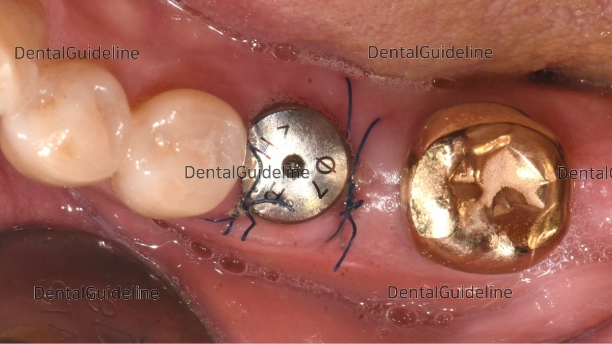
2weeks after the placement.
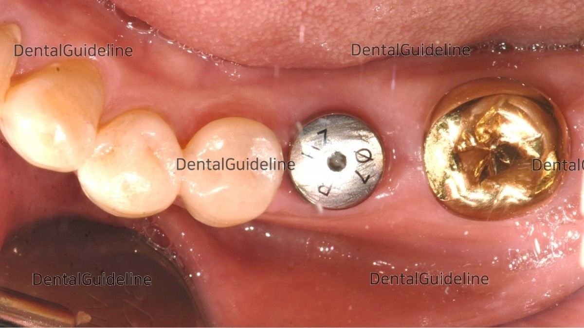
10weeks post-op. intraoral photo on the day of ISQ measurement and impression taking.
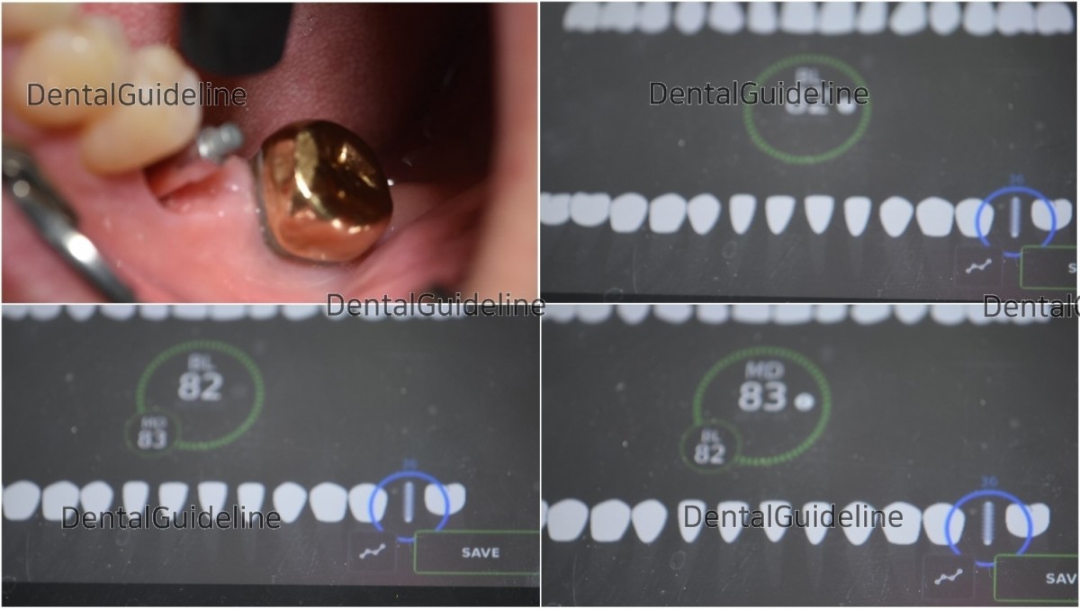
ISQ reading.

custom abutment

orientation jig (=seating jig).

Zirconia crown.
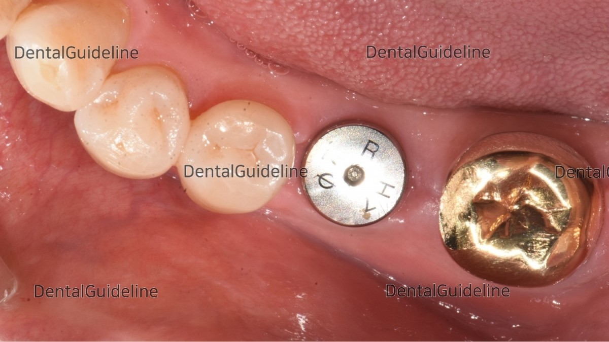
Intra-oral photo on the day of restoration.
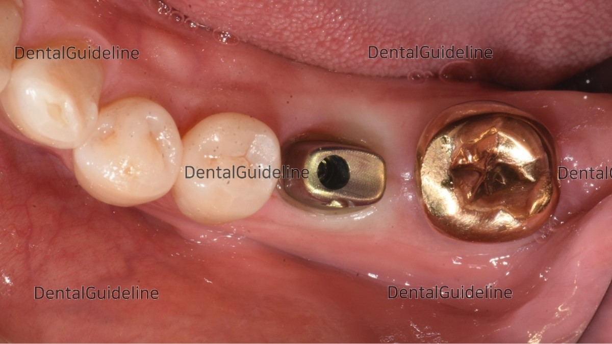
The abutment was connected to the fixture.
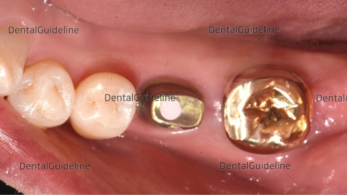
The screw hole was filled with temporary filling material for retrievability.
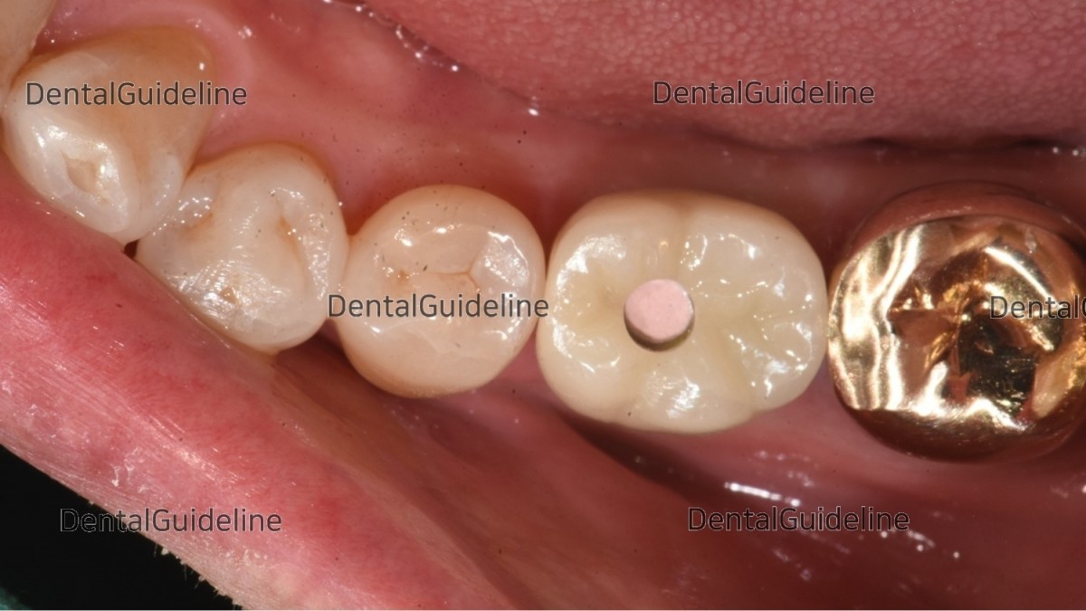
A zirconia crown was seated by adjustment of contact and occlusal surface.
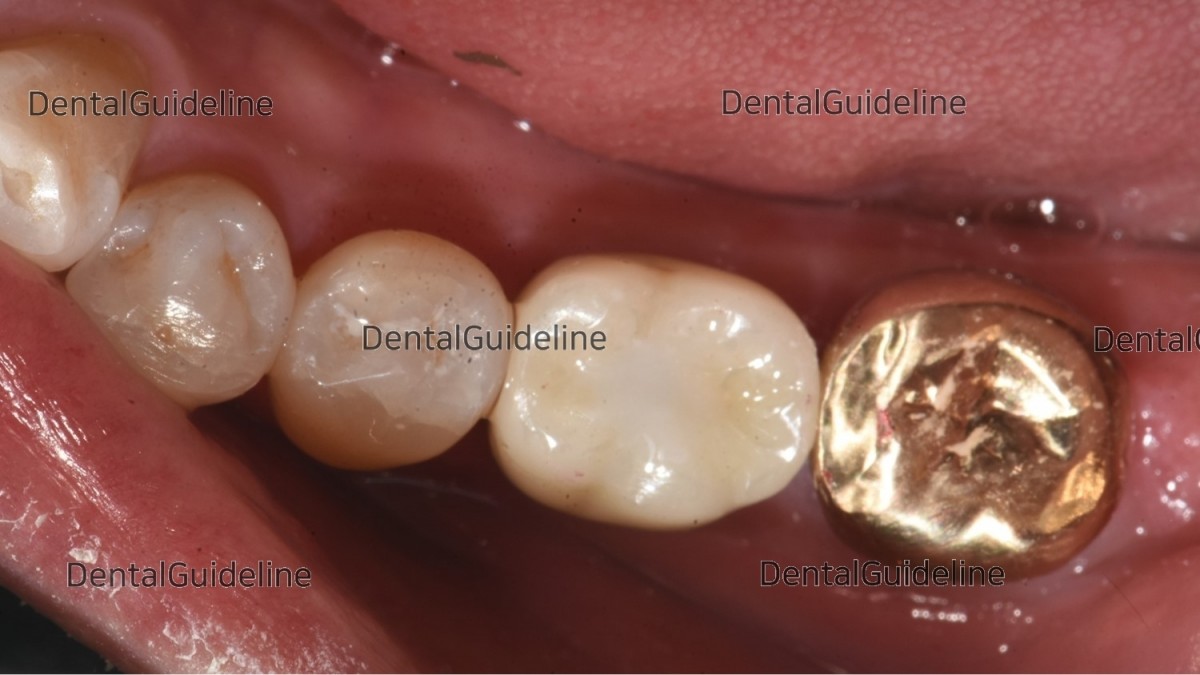
The crown was cemented and the access hole was filled with composite resin.
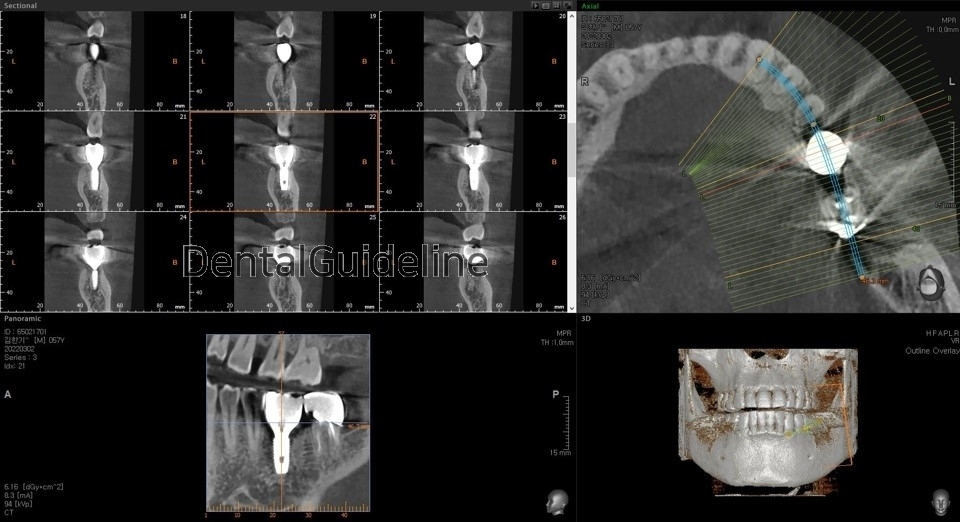
CBCT scan image after crown delivery.
The surgery video will be uploaded sooner or later.
0
- PrevApical surgery. ApicoectomyDec, 12, 2022
- Next7-year follow-up, Immediate placement & GBR, Open membrane method, Dec, 12, 2022
There are no registered comment.


