Implantology
Dental Restoration
Cosmetic Dentistry
Digital Dentistry
Dental Labrotary
Anterior implant placement with Arum implant system in the maxilla.

HAPPYTOGETHER
Views : 3,628/ Jan, 11, 2023
Views : 3,628/ Jan, 11, 2023
<GCjoy>
A 46-year-old female patient didn’t
have any systemic problems but a poor oral condition.
The patient is scheduled
for the implant and general prosthetic restoration in various parts. First of
all, the vertical stop is completed by several prostheses in the posterior
region, and the final stage of intraoral restoration is to proceed with an
anterior implant installation.
 ▲Pre-op intraoral photos.
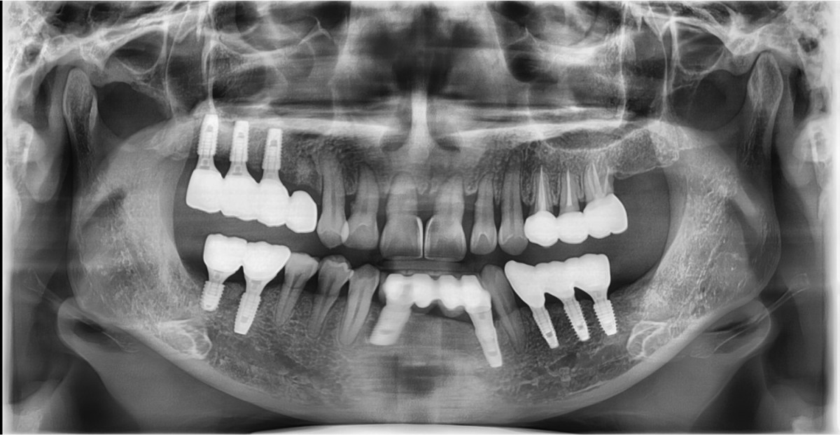 ▲Pre-op panoramic radiograph.
 ▲Extraction
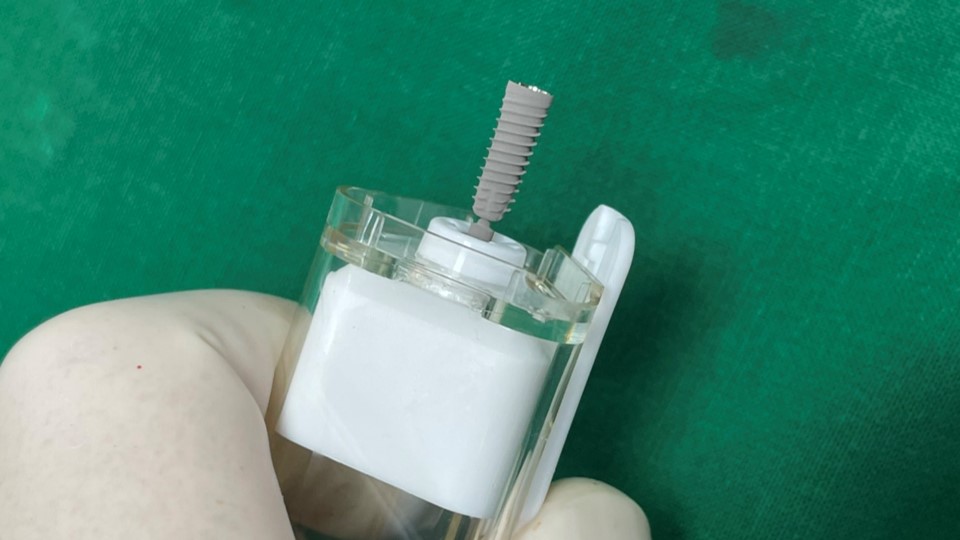 ▲Fixture to be placed. Arum® implant NB1
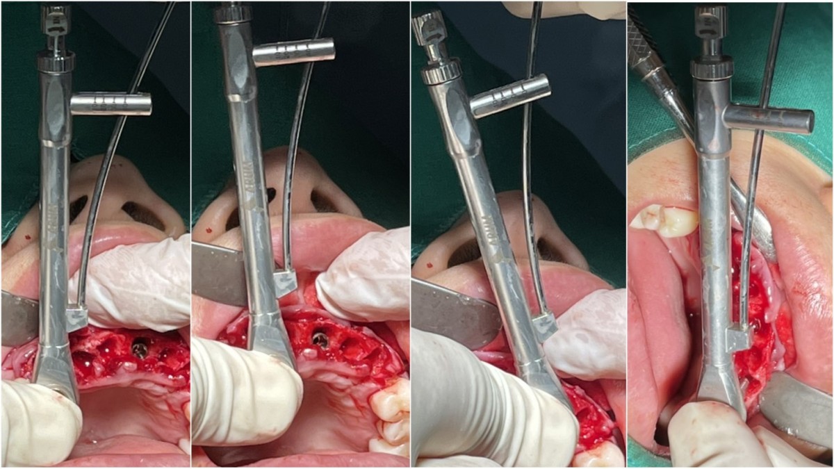 ▲Initial stability value for each implant Arum Dentistry NB1 implant system. Right and left canine 4*11.5, right lateral incisor and left central incisor 4.0*10
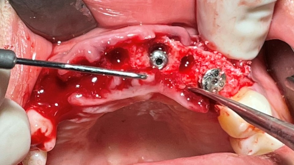 ▲Implant placement GBR
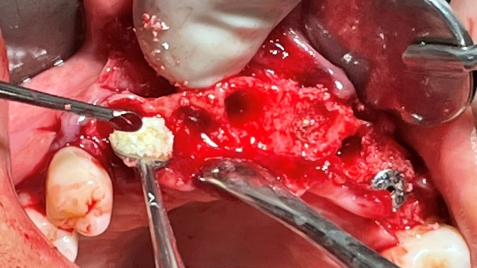 ▲GBR(Xenograft).
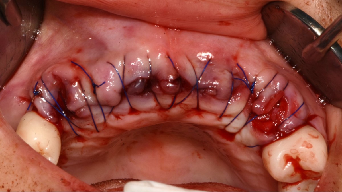 ▲Suture.
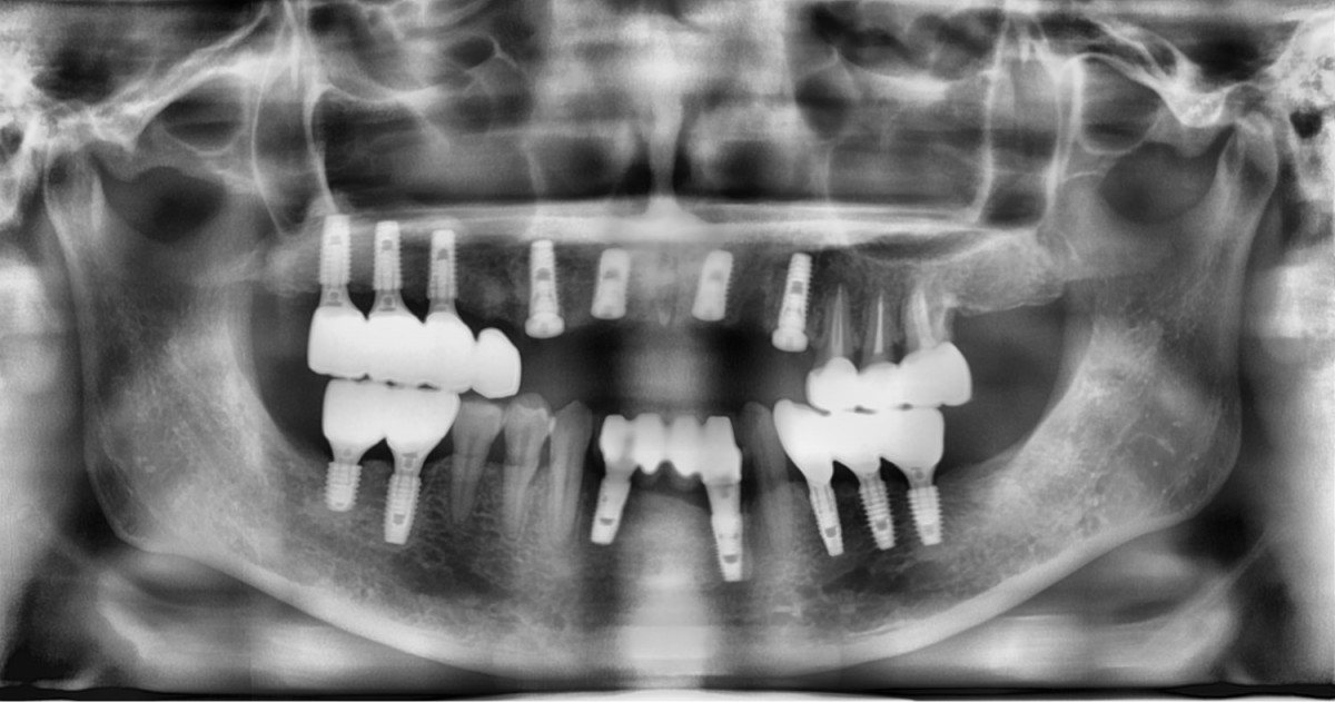 ▲Post-op panoramic radiograph
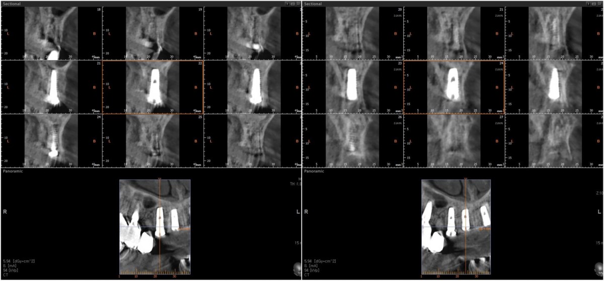 ▲CBCT scan image focused on the canine and lateral incisor on the right side
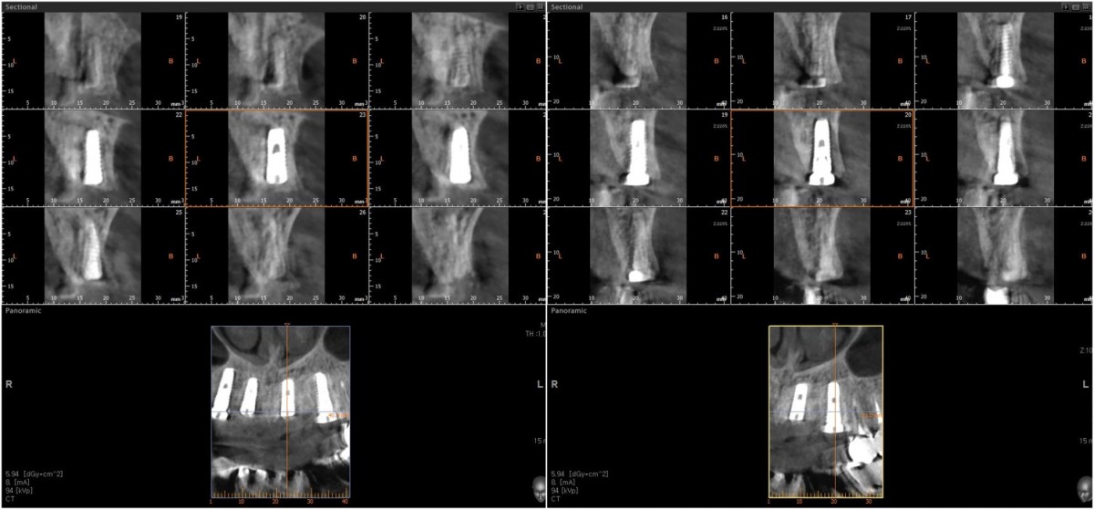 ▲CBCT scan image focused on the central incisor and canine on the left side.
 ▲1 week post-op. HAs were connected immediately in both canine areas.
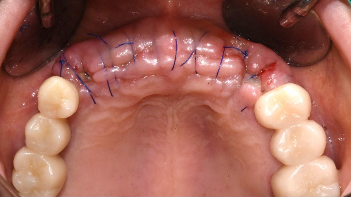
 ▲2 weeks post-op, intraoral photo on the day of suture removal.
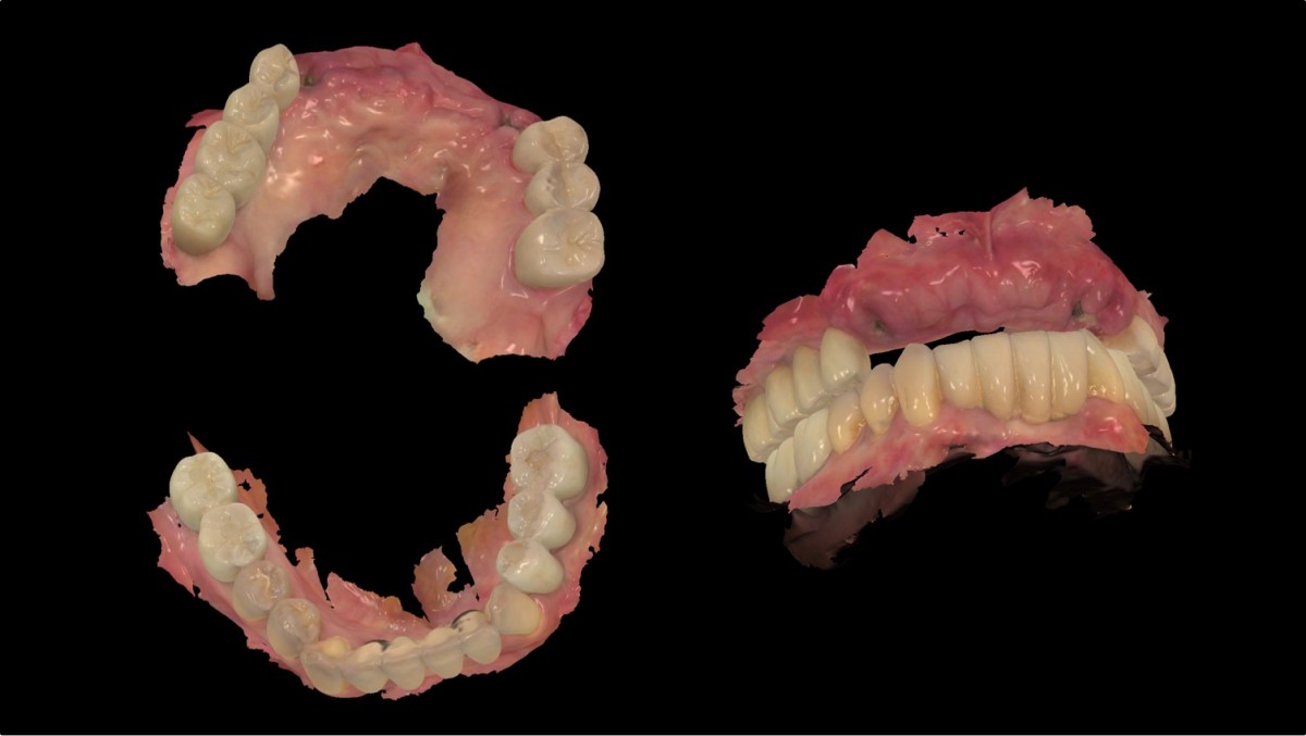 ▲Intraoral scanning for the fabrication of provisional restorations right after suture removal
 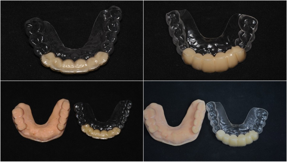 ▲Removable provisional restoration.
 ▲3 weeks post-op. intraoral photo before the delivery of provisional restoration
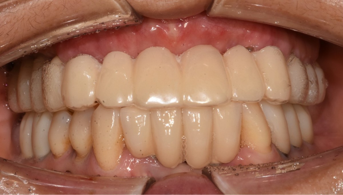 ▲Wearing the appliance of provisional restoration.
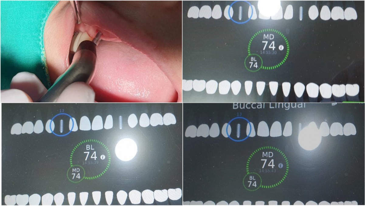 ▲ISQ reading of right canines after 16 weeks of implant placement.
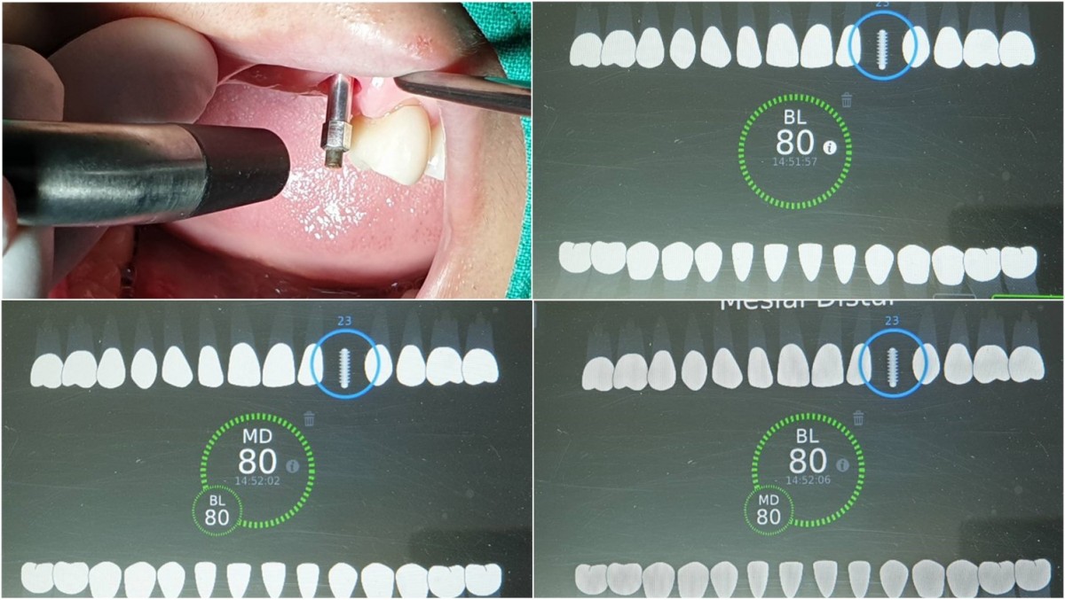 ▲ISQ reading of left canines after 16 weeks of implant placement.
 ▲Intraoral photo before implant uncovery.
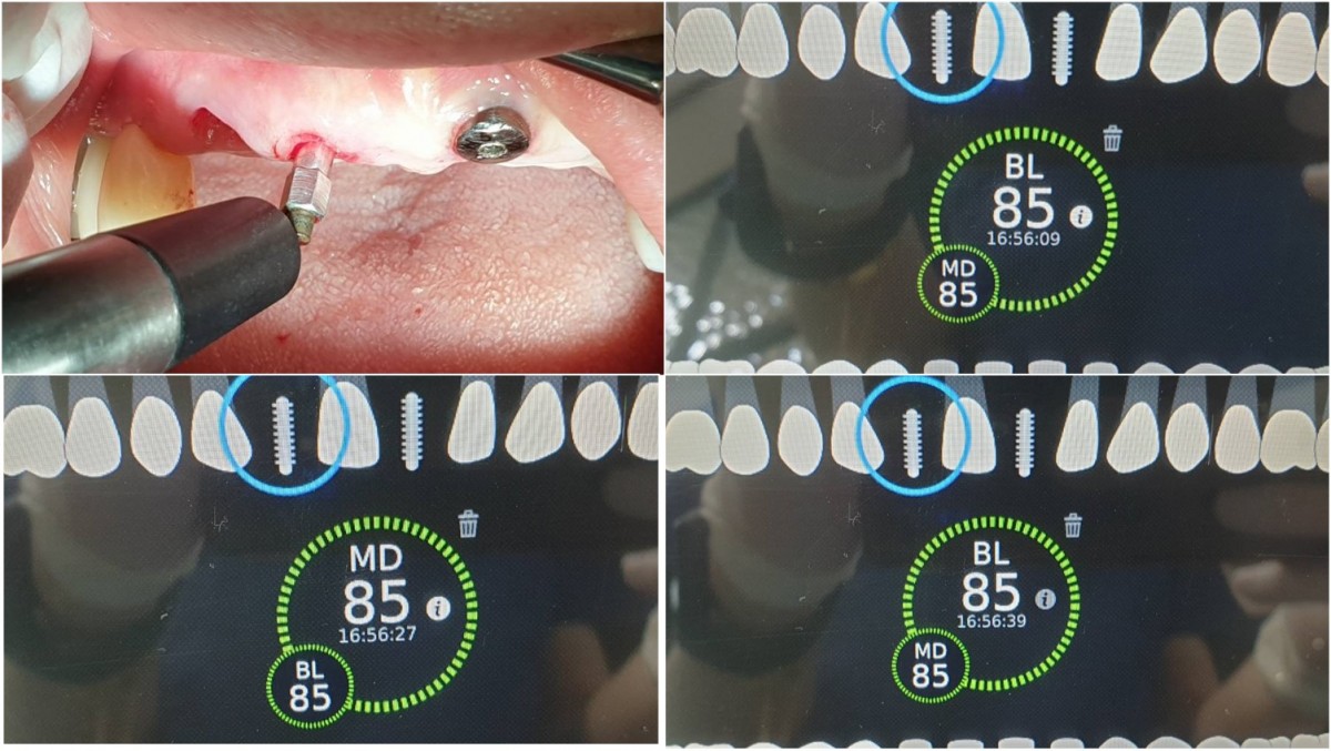 ▲ISQ reading of the fixture in the lateral incisor on the day of 2nd surgery after 18 weeks of implant placement.
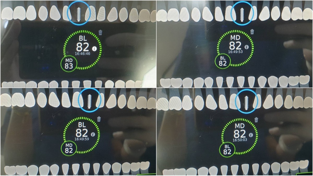 ▲ISQ reading of the fixture in the central incisor
 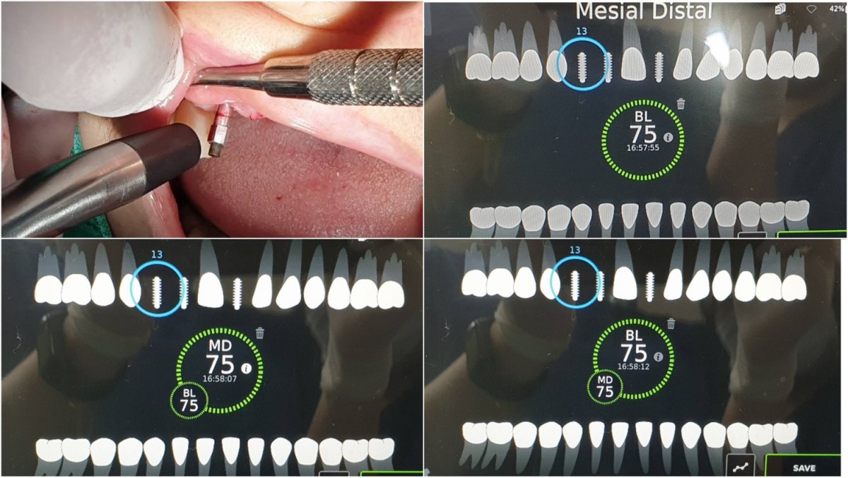 ▲The canine ISQ value was checked again in 2 weeks.
 ▲HA was connected after the tissue punch and ISQ reading.
 ▲Intraoral photo on the day of impression taking.
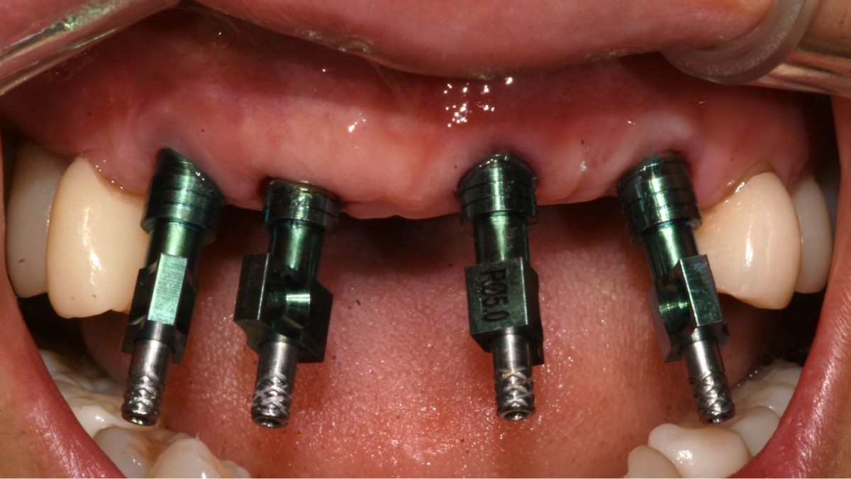 ▲Impression taking
 ▲Customized abutment on the working model
 ▲zirconia prosthesis
 ▲abutments were conneced in the mouth.
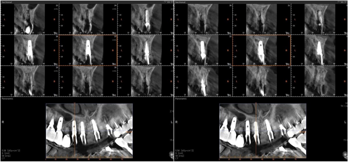 ▲CBCT scan image after abutments connection
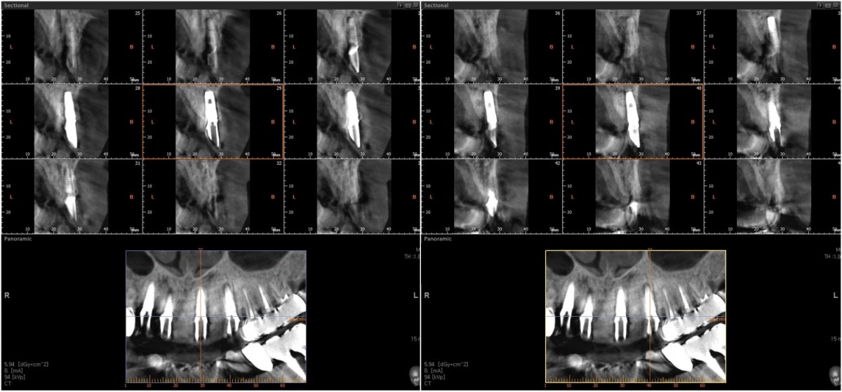 ▲CBCT scan image after abutments connection
 ▲An implant-supported fixed partial denture
0
- PrevImmediate implant placement in the left molar of the maxilla.Jan, 11, 2023
- NextMinor Tooth Movement using tooth positioning appliance Jan, 11, 2023
There are no registered comment.


