Implantology
Dental Restoration
Dental Labrotary
Implant placement after future site development

HAPPYTOGETHER
Views : 3,415/ Dec, 25, 2022
Views : 3,415/ Dec, 25, 2022
<GCkdh> A 40-year-old male had a broken tooth with no symptoms but had discomfort during biting in the adjacent tooth. No systemic issues. 2implants were scheduled after future site development
The socket size of the 1st molar didn’t seem to accept an
immediate placement of the implant, It seemed to get a loss of
initial stability.

Intra-oral view and fractured tooth.
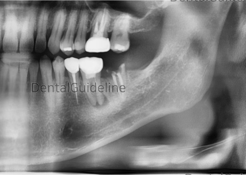
Panoramic radiograph.
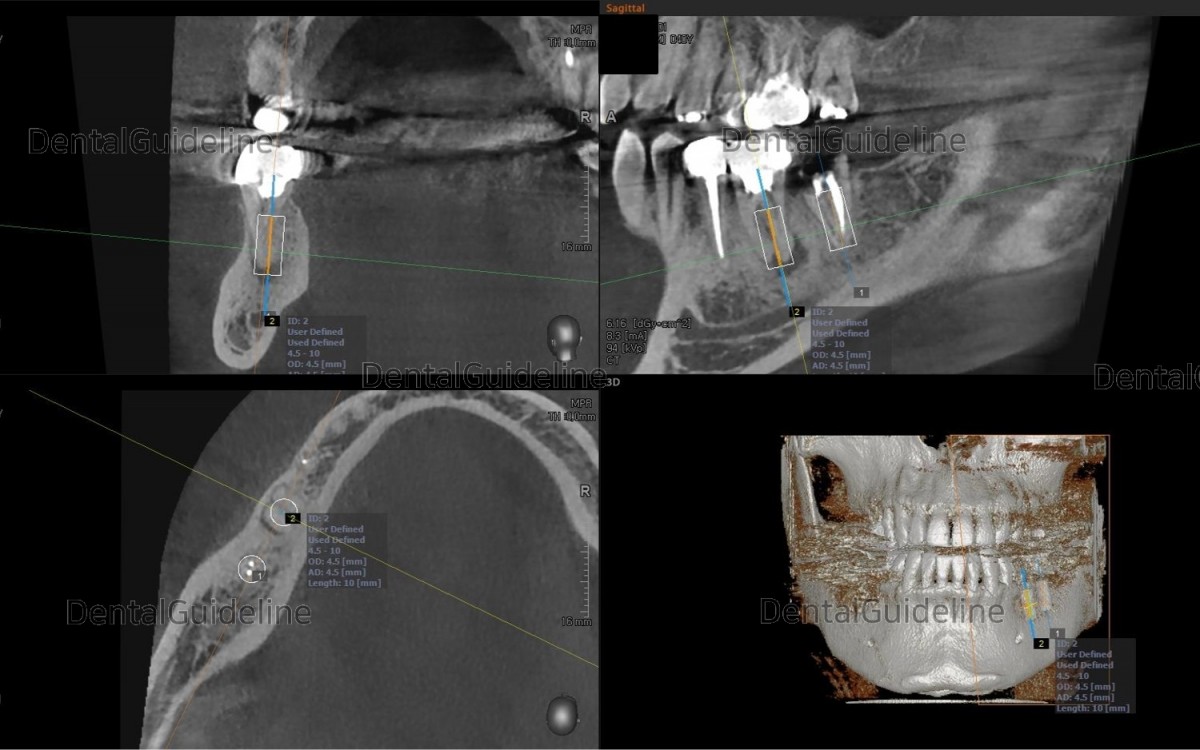
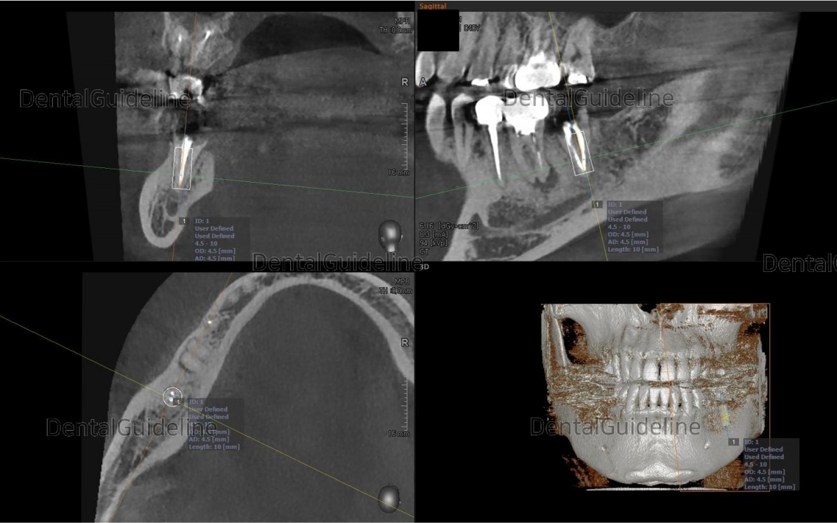
Rough simulation on the CBCT scan views The socket size of the 1st molar didn’t seem to accept the immediate placement of an implant It seems hard to get initial stability.
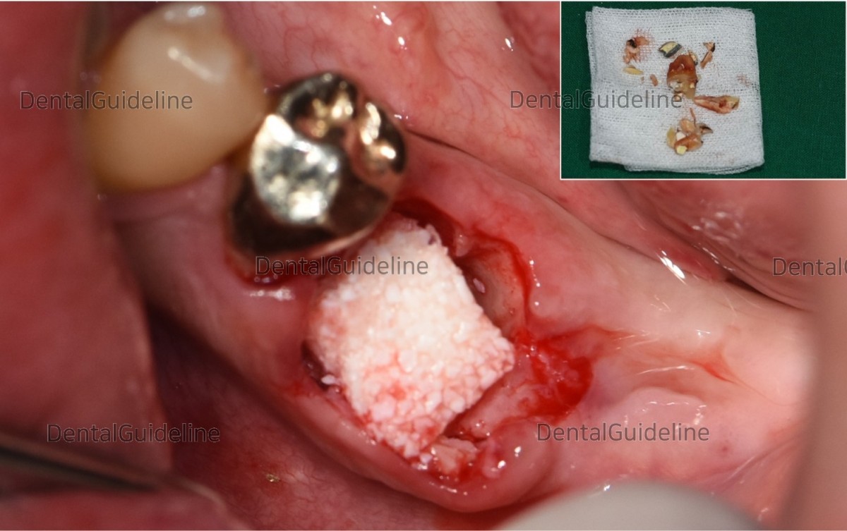
A block grafting material (mixture of HA+collagen) was placed into the socket for future site development.
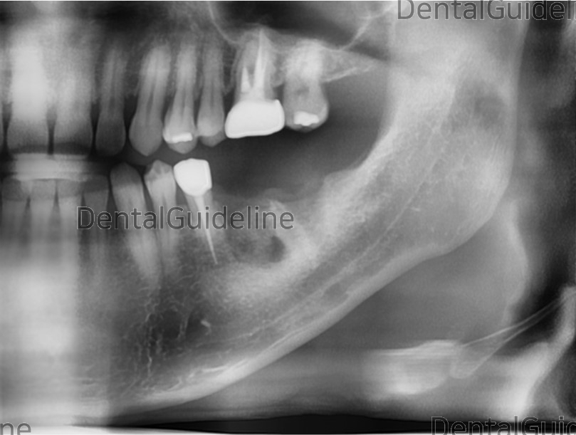
Panoramic radiograph shows future site development in the extraction socket of the 1st molar.
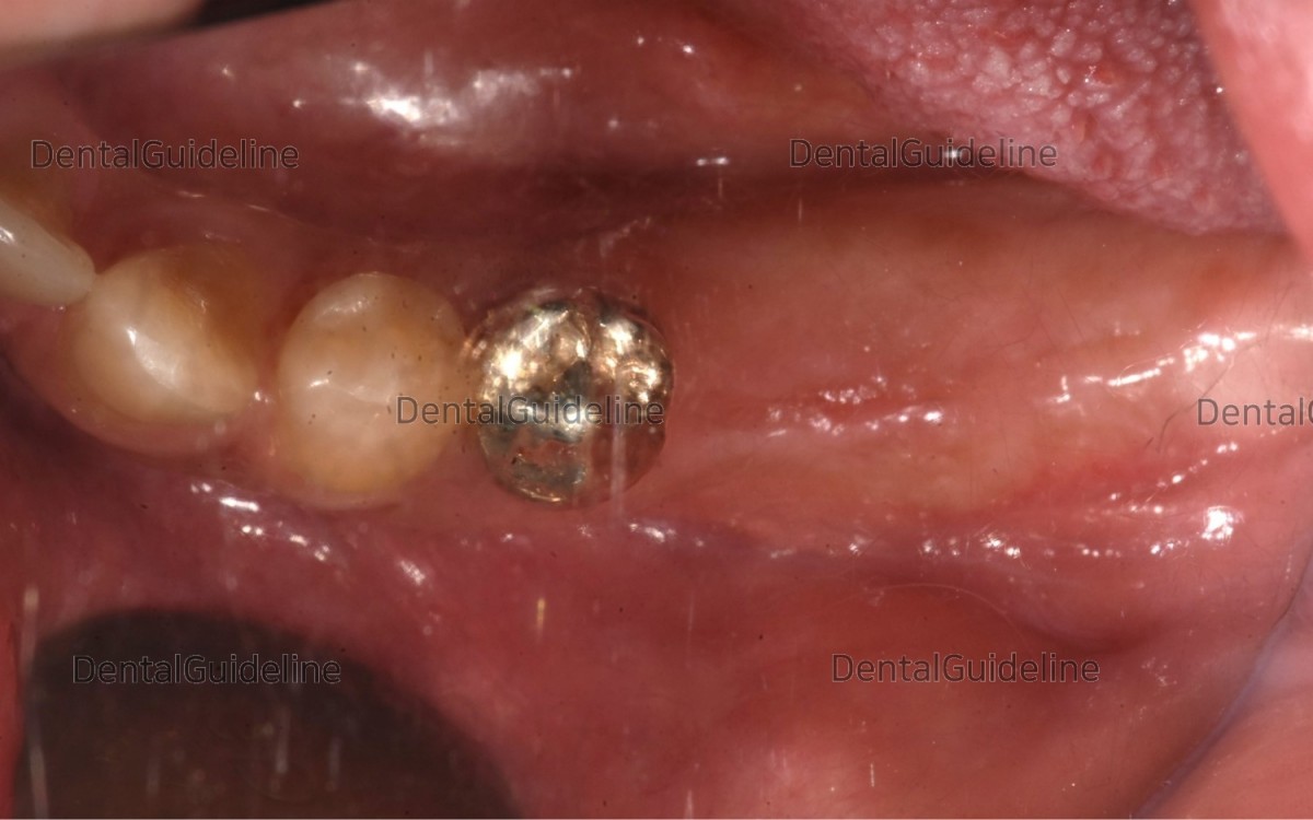
Intra-oral view on the day of implant placement (13 weeks after future site development).
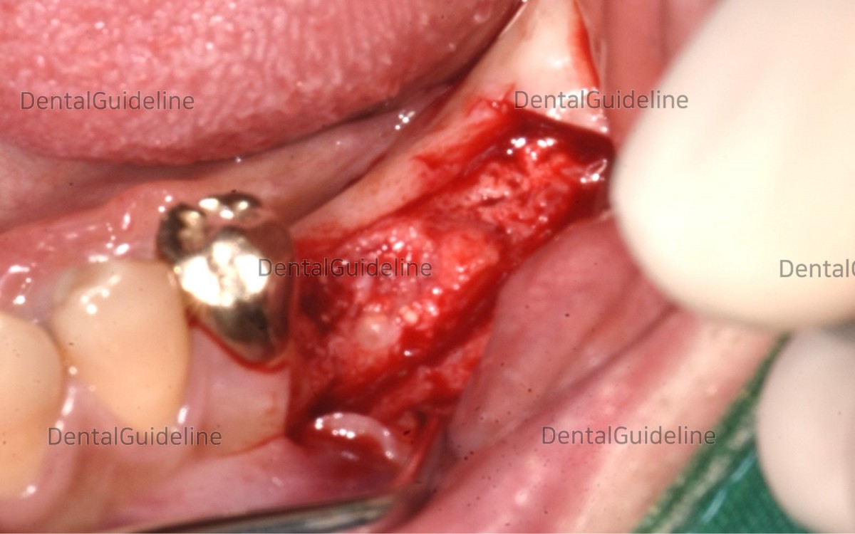
Flap opening. There shows some granulation tissue in the 1st molar area.
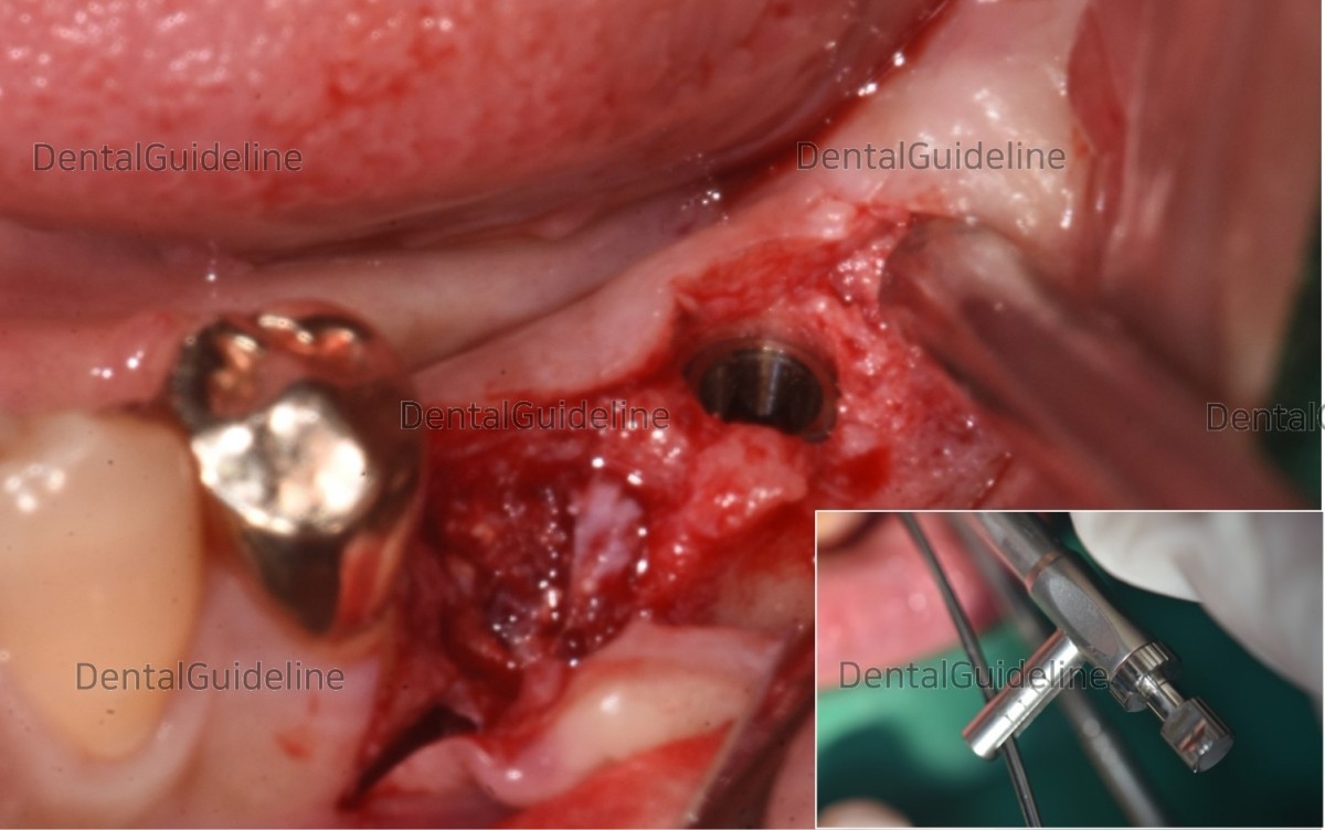
After serial drilling, an Arum Dentistry® Ø4.5/L10mm-sized implant was placed in the 2nd molar zone and had good initial stability(45Ncm).
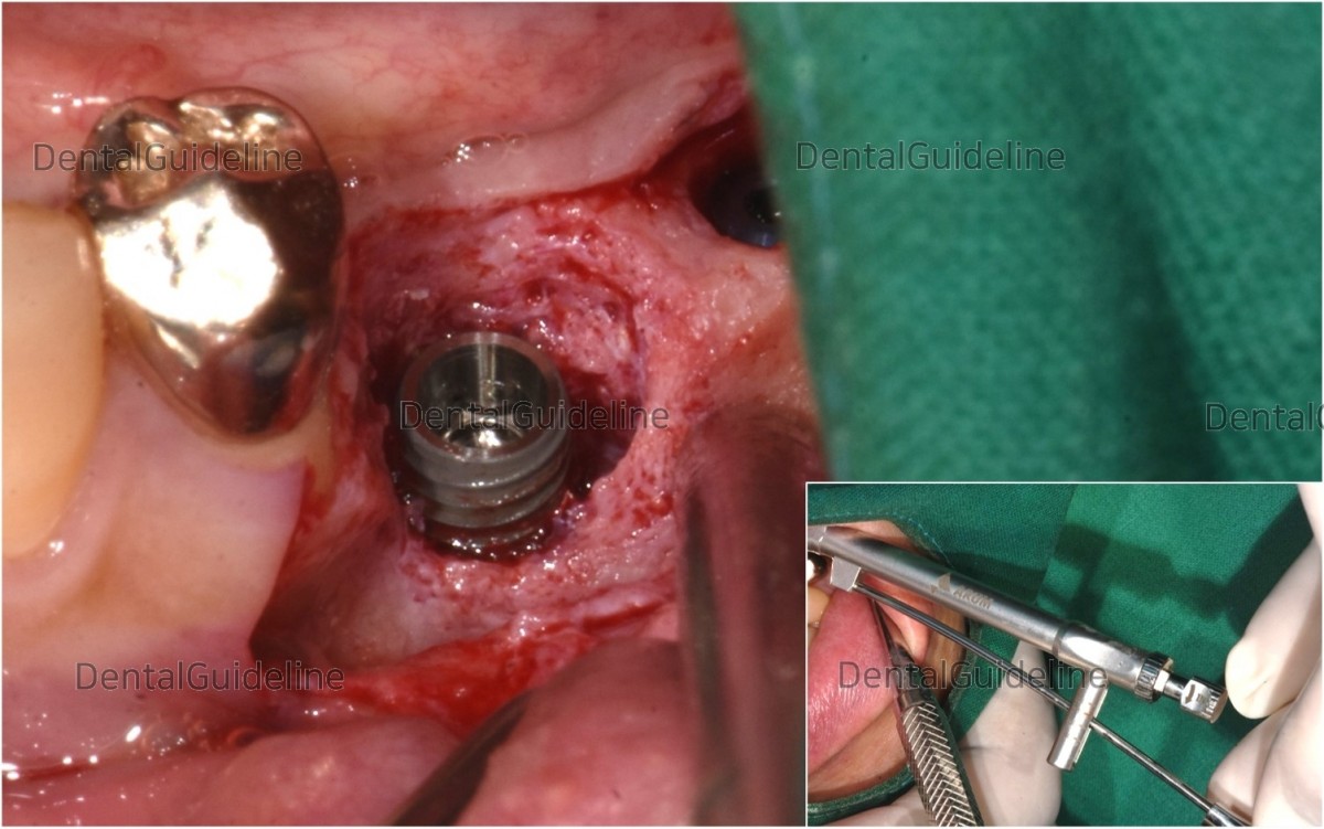
All the existing granulation tissue was removed and an Arum Dentistry® Ø4.5/L10mm-sized implant was placed. The implant in the 1st molar zone also had relatively good initial stability(25Ncm).
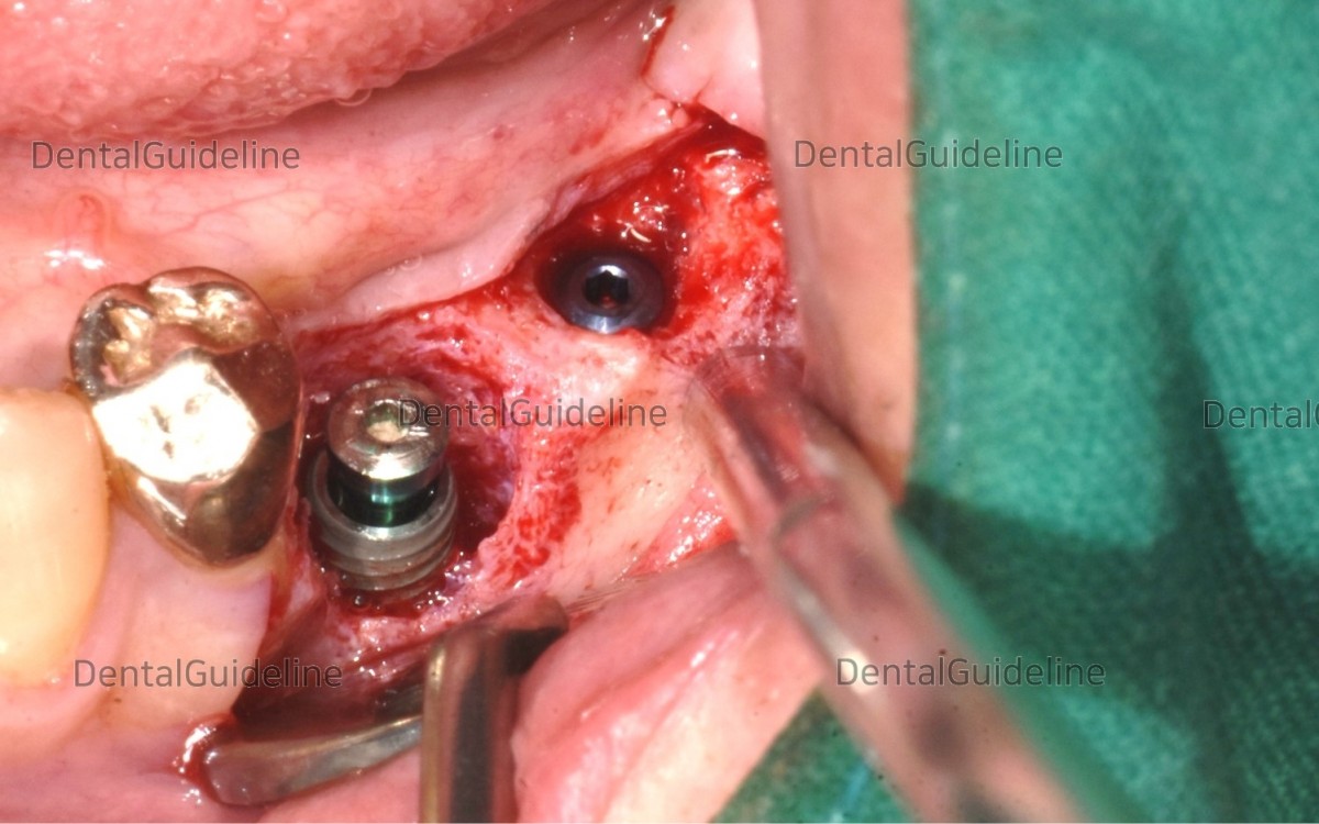
Different types of cover screws were engaged to fixtures. The higher type of cover screw works as a space keeper, which may result in successful GBR.
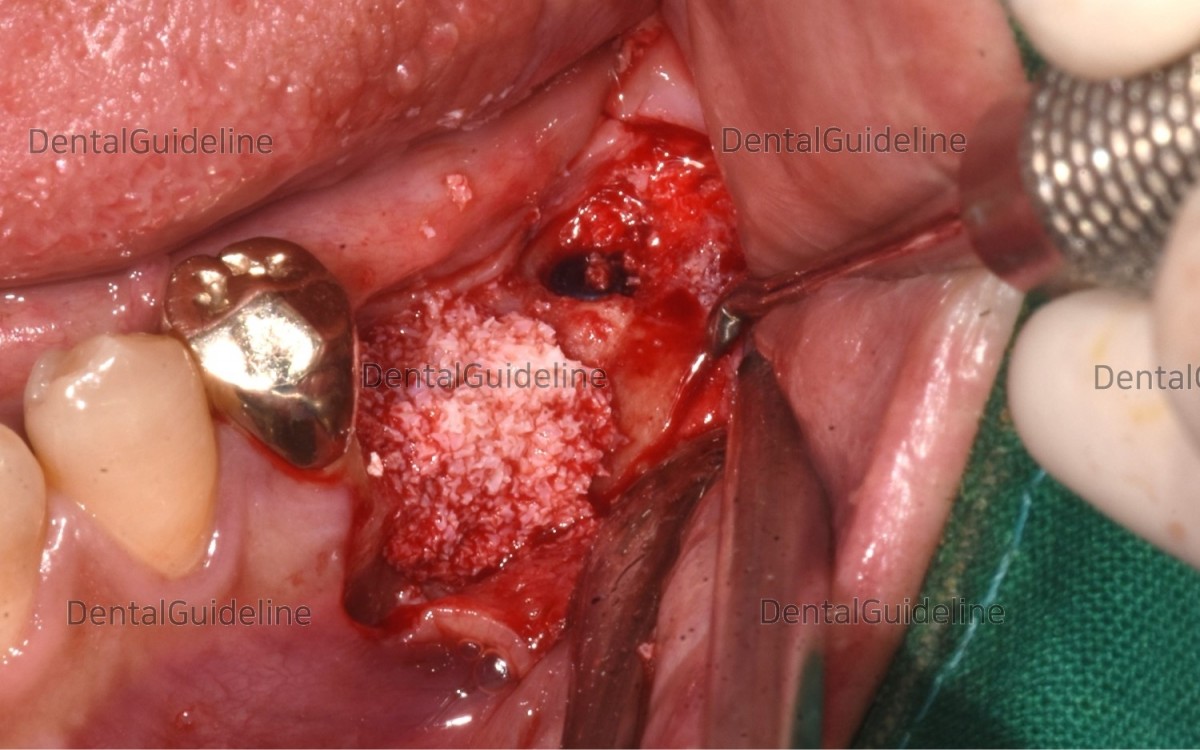
The choice of grafting material(Xeno) was covered around the defective site.
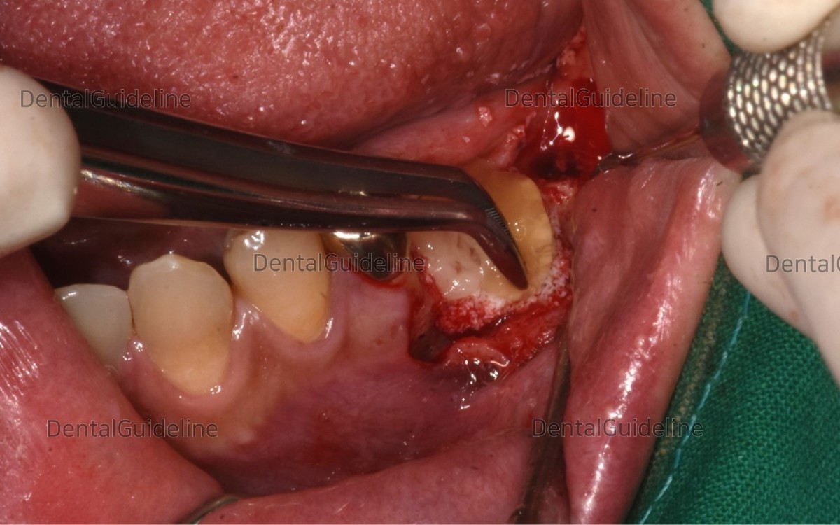
CGF(Concentrated Growth Factor) was laid on the grafting material.
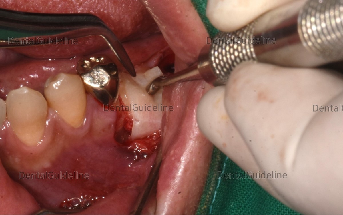
The grafted area was covered with a collagen membrane.
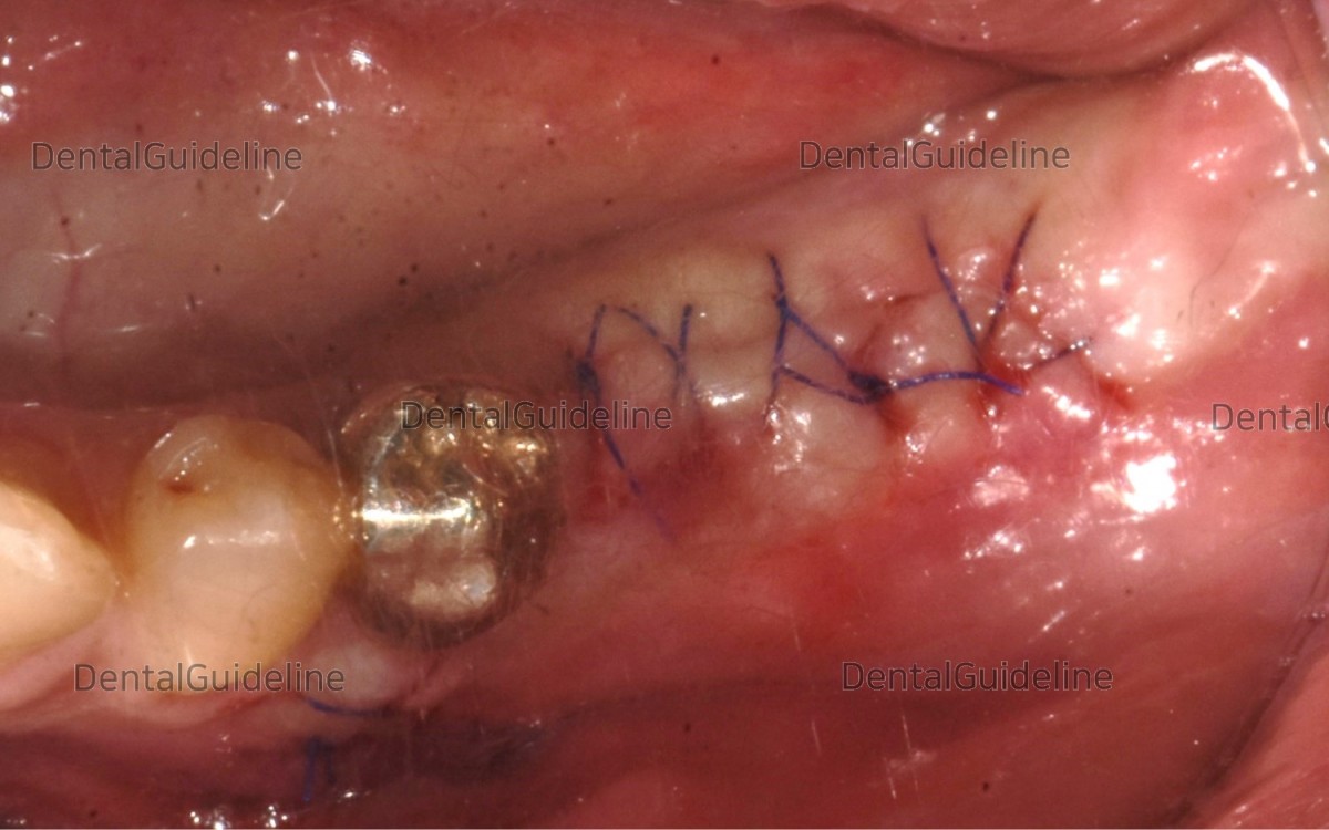
The flap was closed.
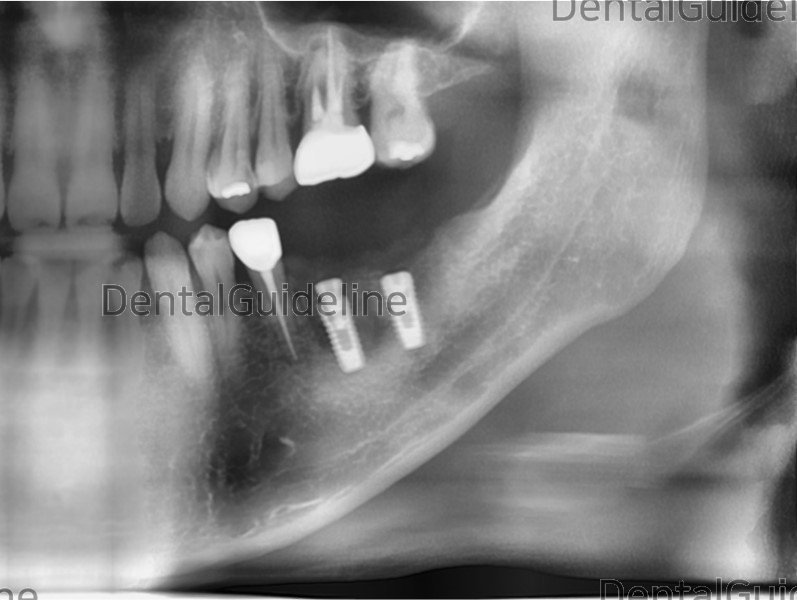
Post-op panoramic radiograph.
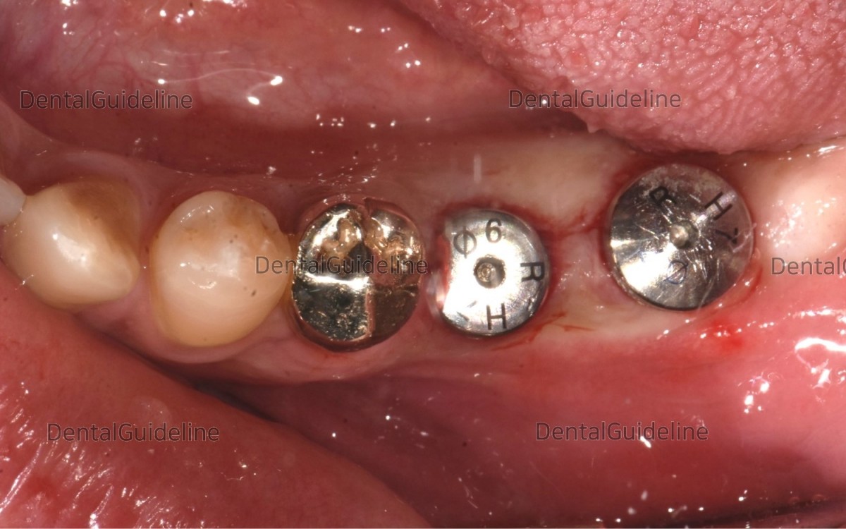
Implant uncovery utilizing the rotary tissue-punching instrument.
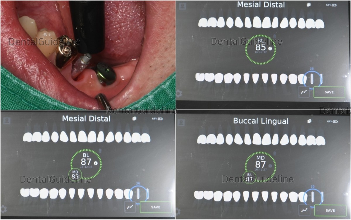
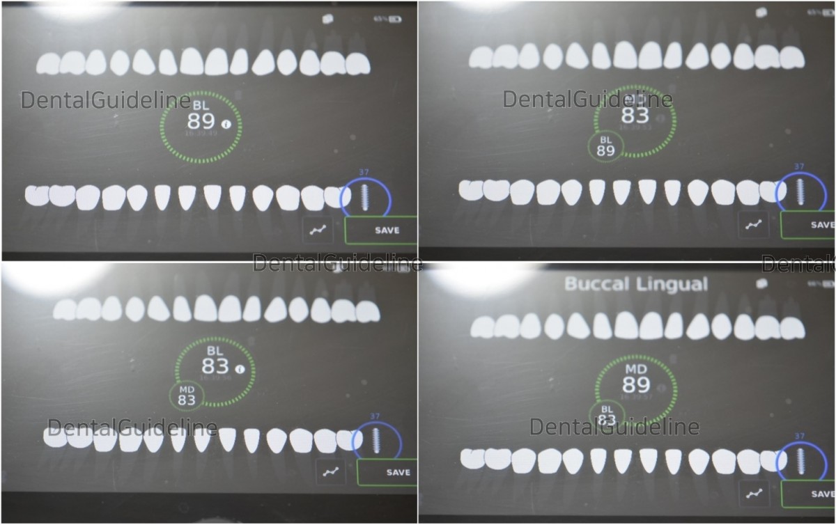
2 weeks after implant uncovery. The ISQ value was good enough at the implant in the molars.
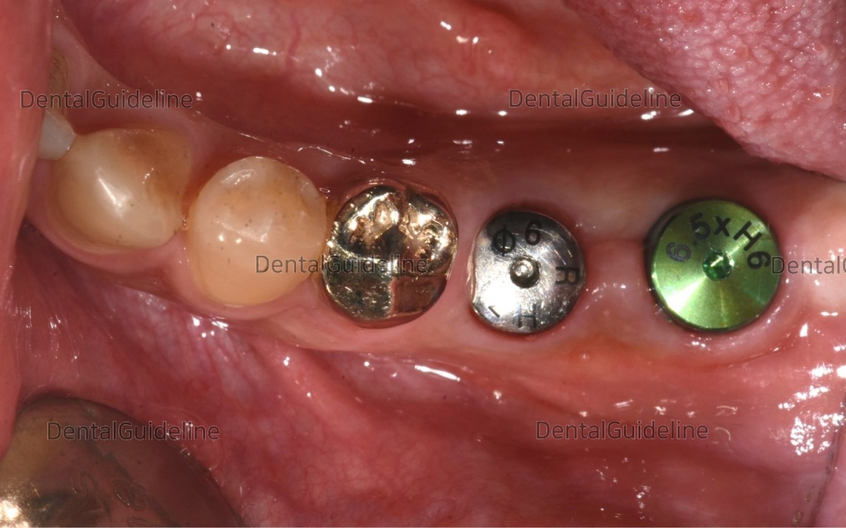
After measurement of ISQ value, Healing abutment was exchanged in needed.
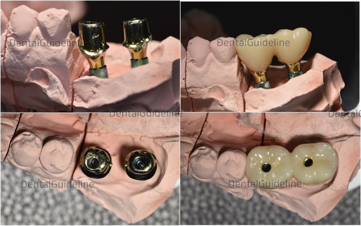
After taking an impression, customized abutments and crowns were fabricated by the dental lab.
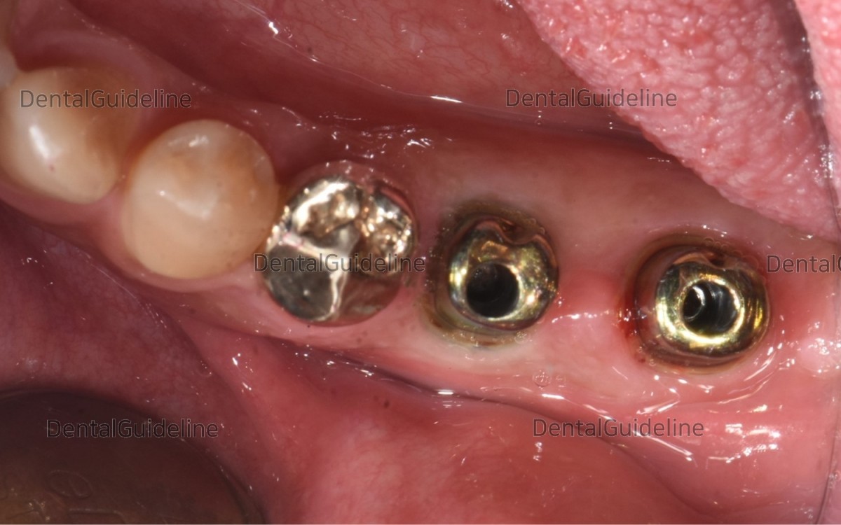
Abutments were connected to the fixture.

The pick-up impression was taken in the condition of crown seating in the mouth. The lab was asked for ceramic to be added on the occlusal surface due to insufficient occlusal contact with opposing dentition.
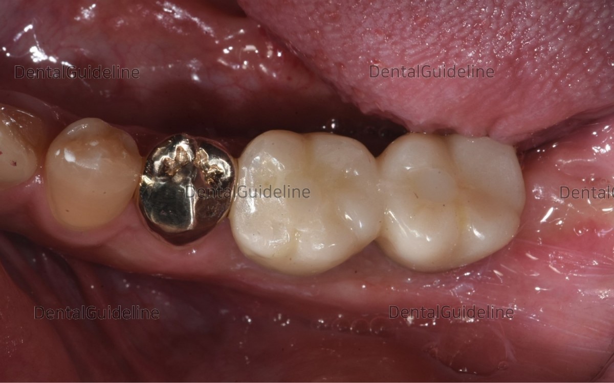
Final cementation and hole filling with composite resin.
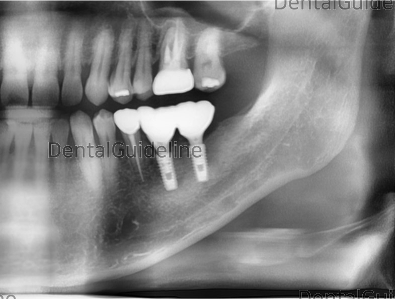
Panoramic radiograph after crown delivery.
0
- PrevCrowns in the esthetic zone.Dec, 25, 2022
- NextPremolar, immediate implant placement Dec, 25, 2022
There are no registered comment.


