Endodontics
Oral Surgery
Apical surgery. Apicoectomy

HAPPYTOGETHER
Views : 1,380/ Dec, 13, 2022
Views : 1,380/ Dec, 13, 2022
<GCkbs> Recurrent fistula and intermittent dull pain in the apical lesion of the canine.
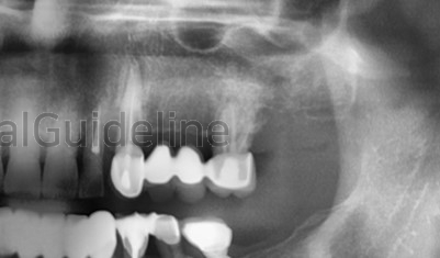
panoramic radiograph

CBCT
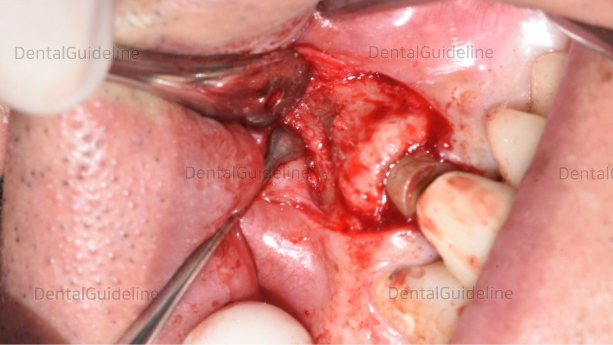
flap opening, lesion exposure.

The apical part was cut using a high-speed bur and the granulation tissue was curette out.
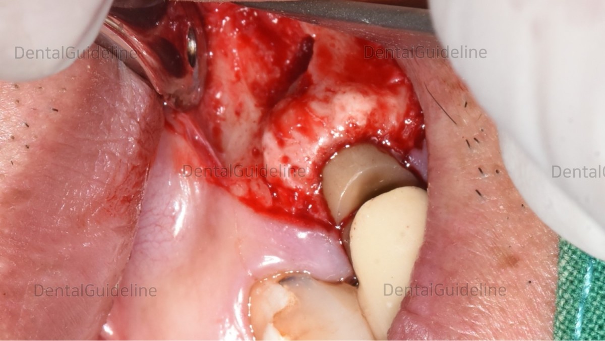
The cut site of the root was filled with MTA (Mineral Trioxide Aggregate).
(It is not visible in this photo due to the shooting angle).
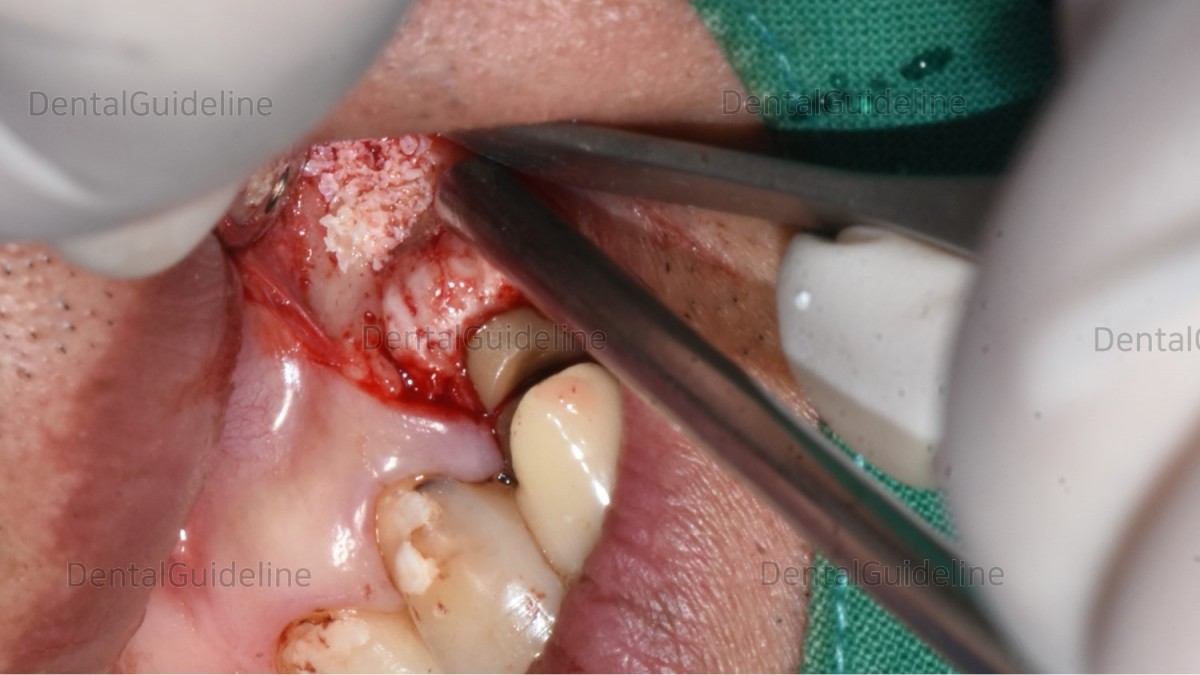
After that, GBR was performed.
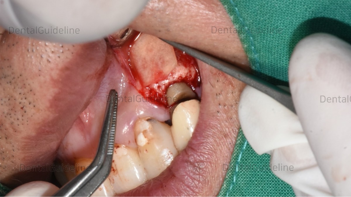
collagen membrane.

suture.

Panoramic radiograph
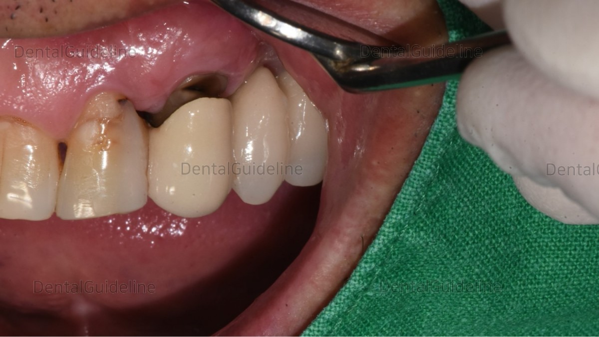
1 month after the apicoectomy.
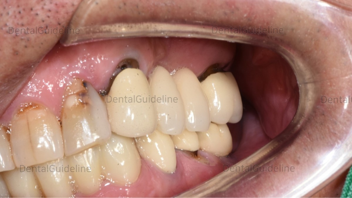
2.5 years later
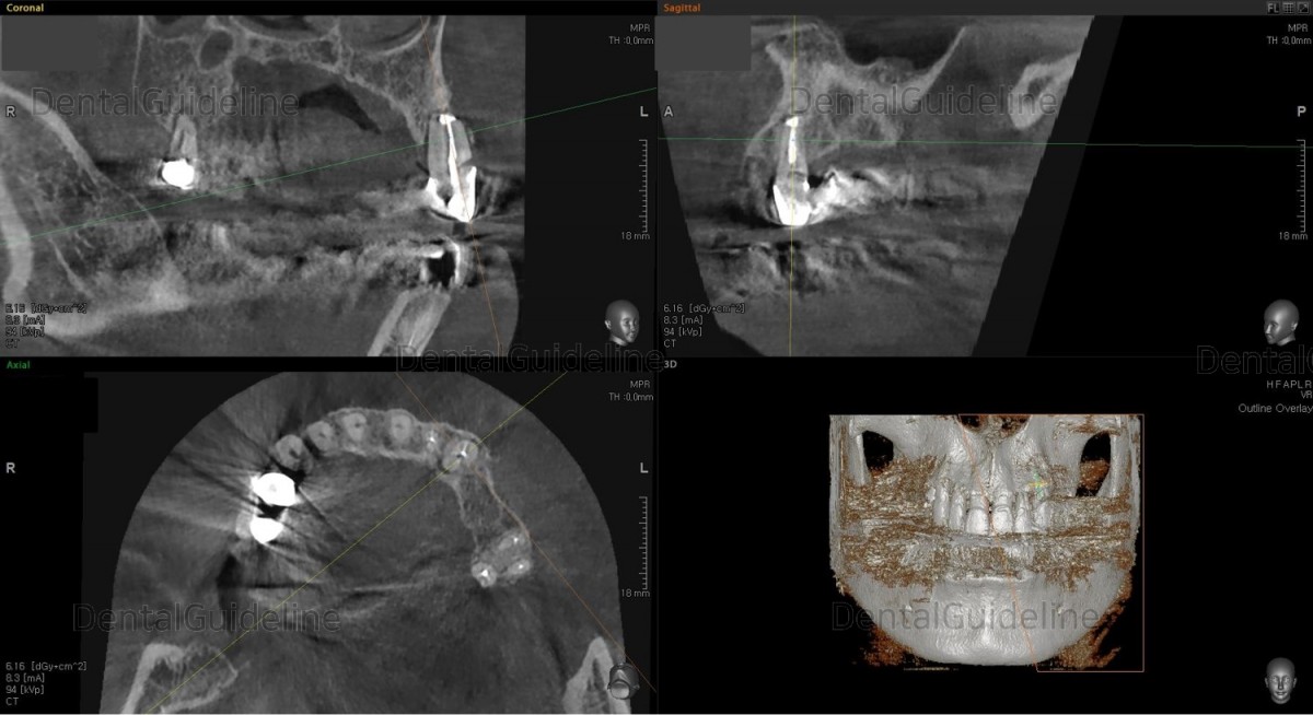
CBCT. 2.5 years of the apicoectomy.

panoramic radiograph

CBCT

flap opening, lesion exposure.

The apical part was cut using a high-speed bur and the granulation tissue was curette out.

The cut site of the root was filled with MTA (Mineral Trioxide Aggregate).
(It is not visible in this photo due to the shooting angle).

After that, GBR was performed.

collagen membrane.

suture.

Panoramic radiograph

1 month after the apicoectomy.

2.5 years later

CBCT. 2.5 years of the apicoectomy.
0
- PrevShade selection for prosthesisDec, 13, 2022
- NextImmediate placement, GBR, Arum NB-1 Dec, 13, 2022
There are no registered comment.


