Dental Restoration
Endodontics
Periodontics
Crown lengthening & restoration 7-year follow-up

HAPPYTOGETHER
Views : 2,114/ Nov, 29, 2022
Views : 2,114/ Nov, 29, 2022
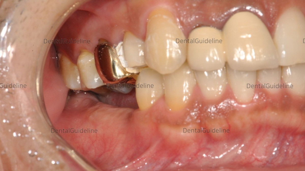
1. Initial photo. Due to the loss of the mandibular teeth for a long period, the maxillary teeth came down. For implant-supported restoration of the edentulous mandibular region, the occlusal elevation of the opposing maxillary teeth is necessary first.
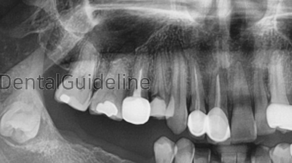
2. Initial panoramic radiograph.
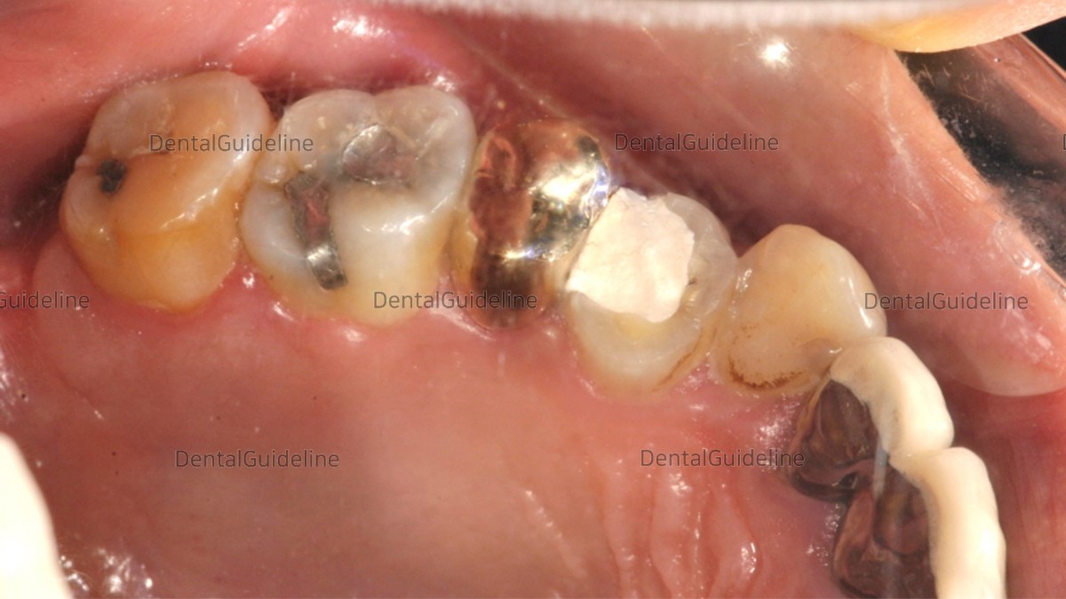
3. Since it did not seem to be possible to secure sufficient interdental space with occlusal surface reduction alone, root canal treatment and crown were decided.
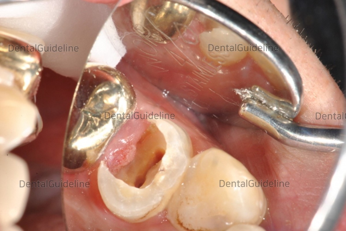
4. A lot of caries has progressed subgingivally, so it is not possible to form a normal prosthetic margin.
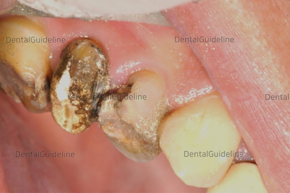
5. In the 2nd premolar, when the old crown was removed, caries progressed a lot.
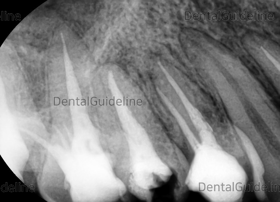
6. root canal treatment was done.
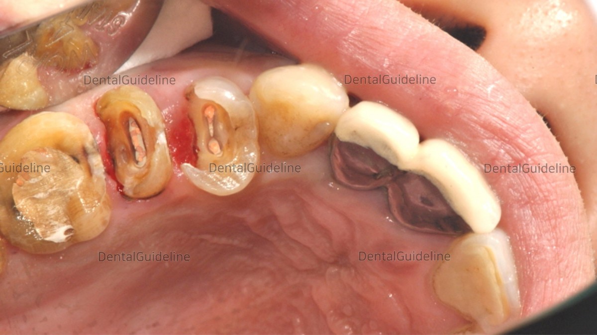
7. A photo of the patient after removing all existing dental caries.
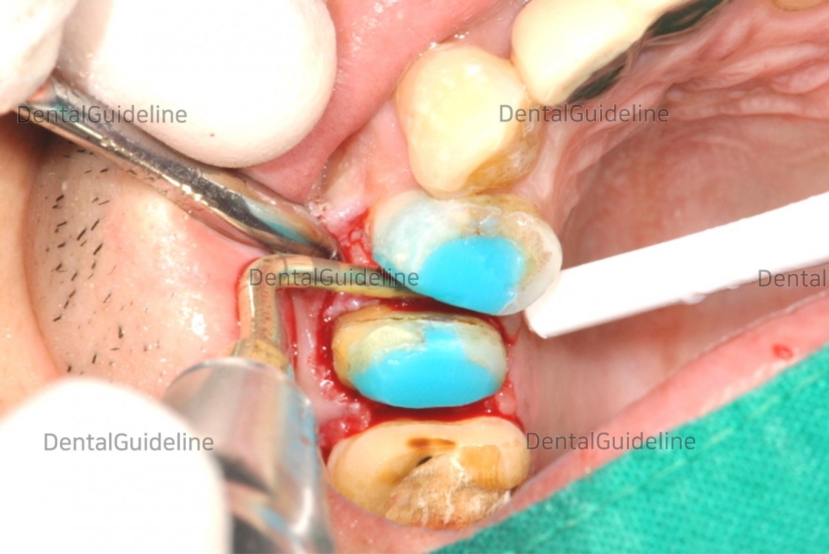
8. Photo of opening the flap and performing crown lengthening surgery.
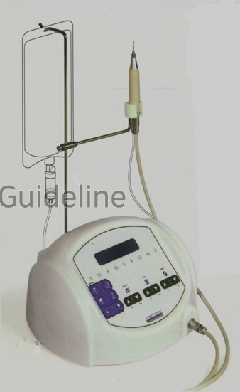
9. piezoelectric surgery machine (SURGYBONE®, SILFRADENT Co.)
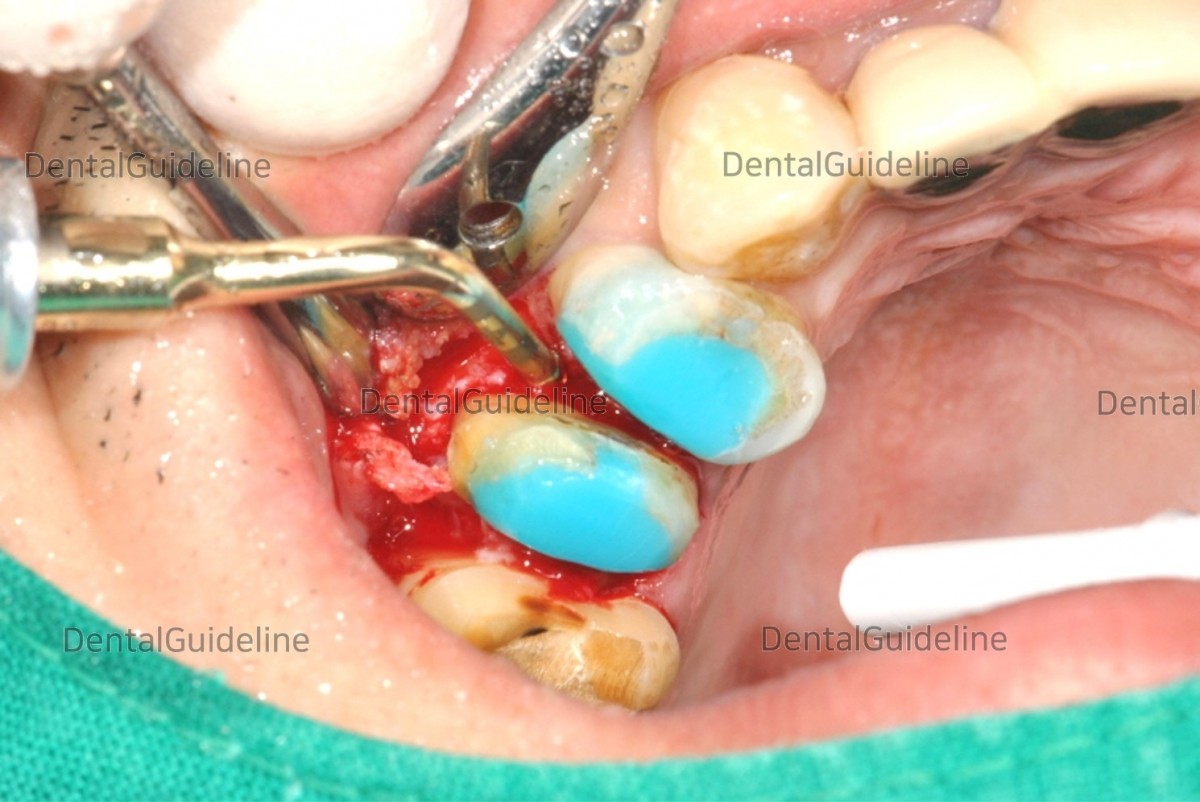
10. A photo of removing the alveolar bone between the teeth and around the teeth using a piezo surgical machine.
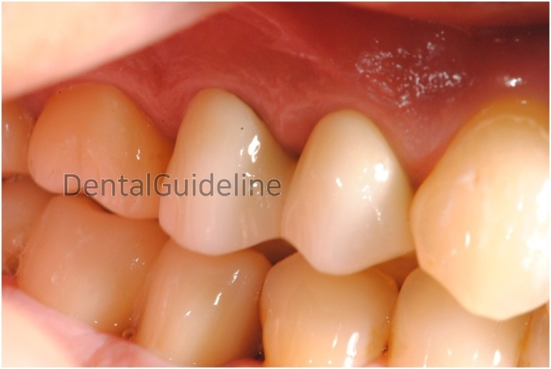
11. Intraoral photo after restoration.
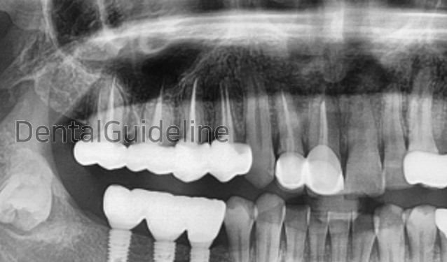
12. panoramic radiograph.
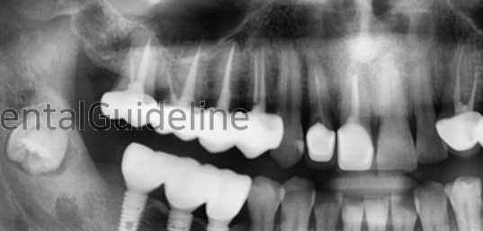
13. panoramic radiograph after 7 years and 4 months.
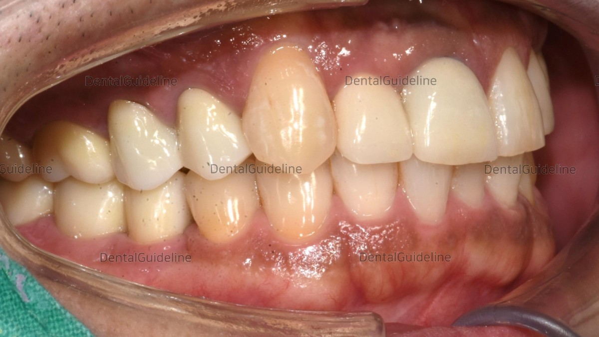
14. Intraoral photo after 7 years and 4 months. (lateral view)
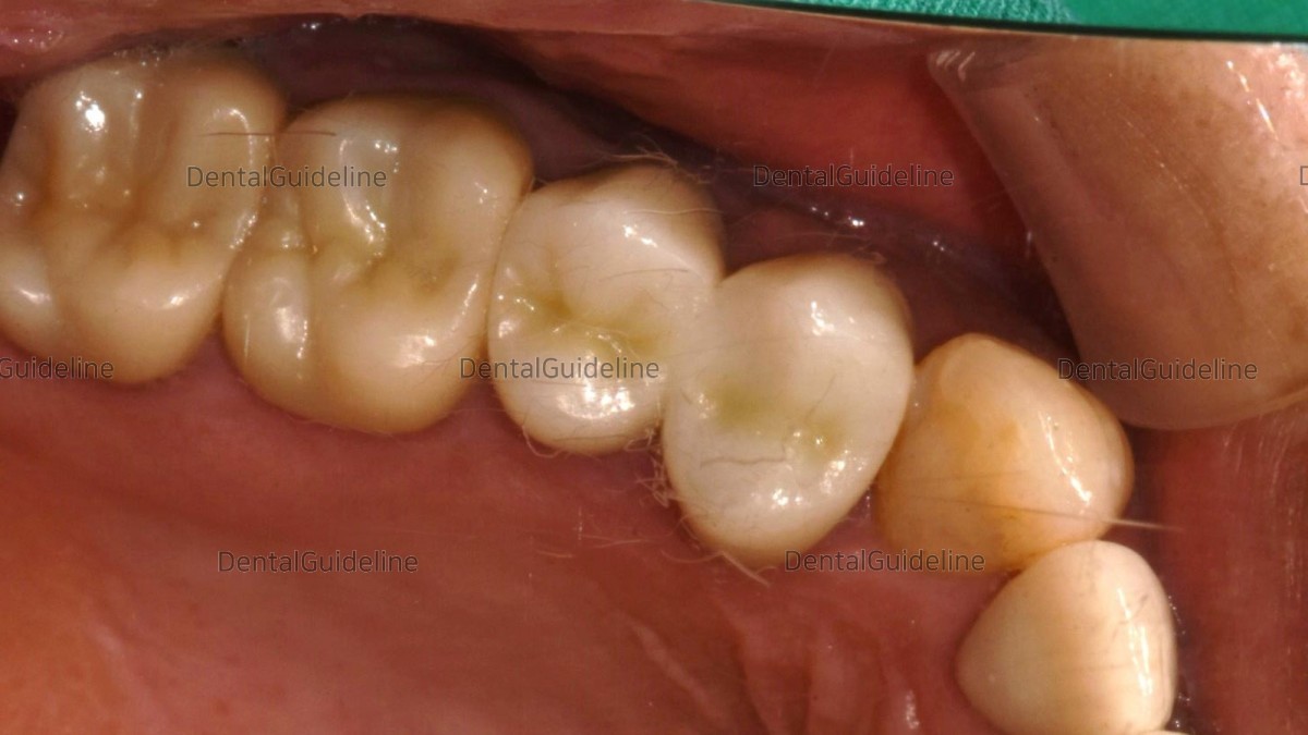
15. Intraoral photo after 7 years and 4 months. (occlusal view)

1. Initial photo. Due to the loss of the mandibular teeth for a long period, the maxillary teeth came down. For implant-supported restoration of the edentulous mandibular region, the occlusal elevation of the opposing maxillary teeth is necessary first.

2. Initial panoramic radiograph.

3. Since it did not seem to be possible to secure sufficient interdental space with occlusal surface reduction alone, root canal treatment and crown were decided.

4. A lot of caries has progressed subgingivally, so it is not possible to form a normal prosthetic margin.

5. In the 2nd premolar, when the old crown was removed, caries progressed a lot.

6. root canal treatment was done.

7. A photo of the patient after removing all existing dental caries.

8. Photo of opening the flap and performing crown lengthening surgery.

9. piezoelectric surgery machine (SURGYBONE®, SILFRADENT Co.)

10. A photo of removing the alveolar bone between the teeth and around the teeth using a piezo surgical machine.

11. Intraoral photo after restoration.

12. panoramic radiograph.

13. panoramic radiograph after 7 years and 4 months.

14. Intraoral photo after 7 years and 4 months. (lateral view)

15. Intraoral photo after 7 years and 4 months. (occlusal view)
0
- PrevPorcelain crowns & shade selection.Nov, 29, 2022
- NextCrown lengthening, 8-year follow-up. Nov, 29, 2022
There are no registered comment.


