Implantology
Dental Restoration
Orthodontics
Digital Dentistry
Maxillary Sinus Graft, Implant, MTM(Minor Tooth Movement), Oral Scanning

HAPPYTOGETHER
Views : 1,281/ Nov, 14, 2022
Views : 1,281/ Nov, 14, 2022
A 60-year-old female patient had
her teeth extracted about half a year ago. She had a cholecystectomy history
and had been taking osteoporosis medications every week.
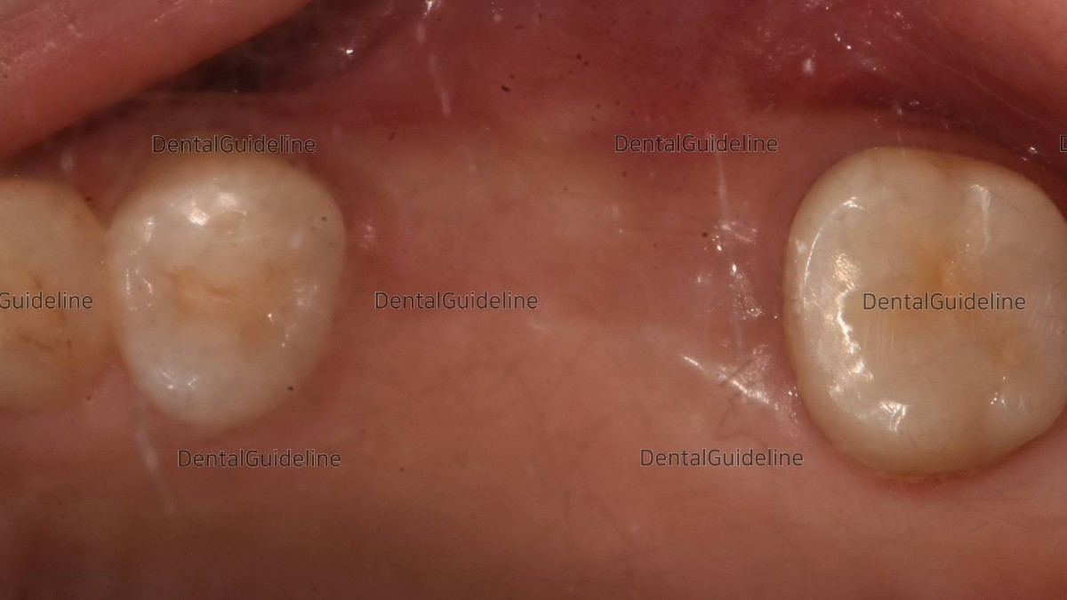
1. Initial photo. Two teeth were extracted about half a year ago.
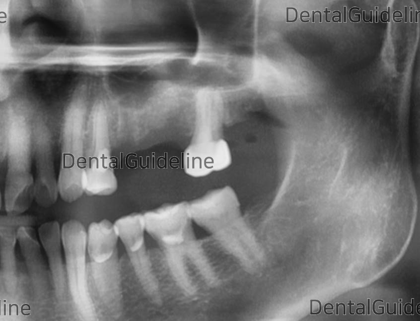
2. panoramic radiograph was taken to check the upper edentulous area.
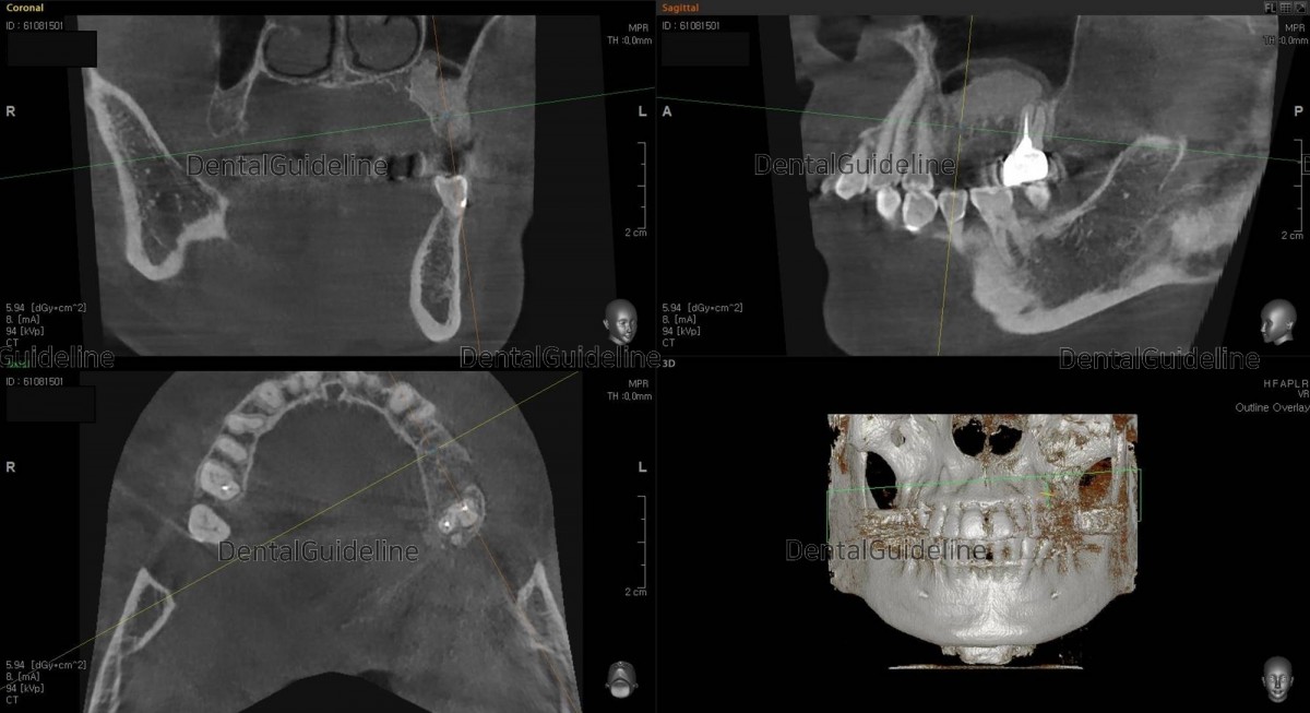
3 CBCT scan image after laterally approached sinus graft.
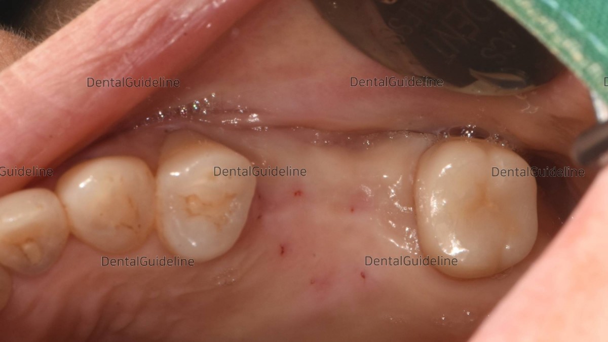
4. Intra-oral photo was taken on the day of implant placement.
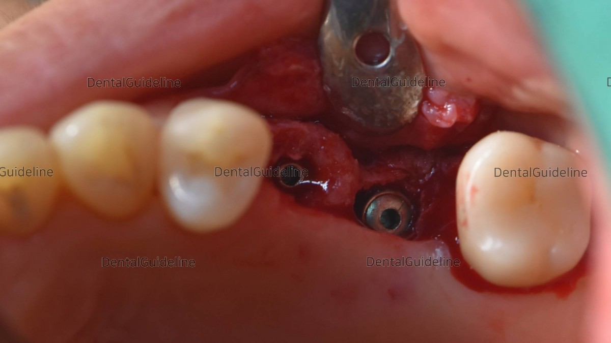
5. 2 implants were placed using a series of ridge expanders - ARUMDENTISTRY Co. NB-1 Ø4/10 (30Ncm) at the premolar zone and NB-1 Ø5/10 (20Ncm) at the molar zone
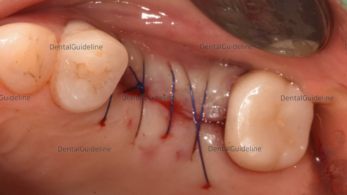
6. Slight GBR was performed according to conventional GBR protocol and the flap was closed.
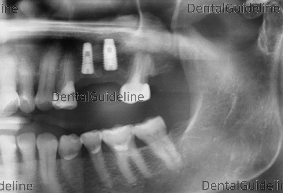
7. Panoramic radiograph right after implant surgery.
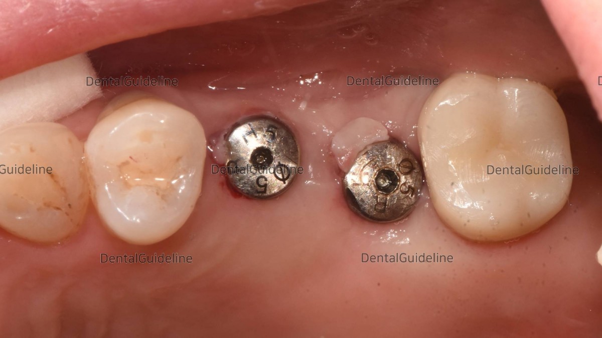
8. Gingival tissue punching was performed to expose fixtures. The 2nd molar showed unexpected mesial movement during the osseointegration period of the implant.
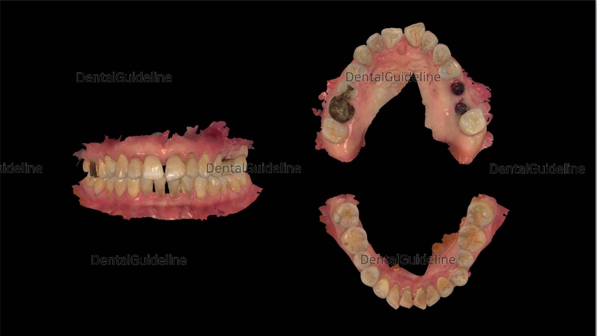
9. Intra-oral scanning was done to fabricate a tooth-positioning appliance for the 2nd molar distalization.
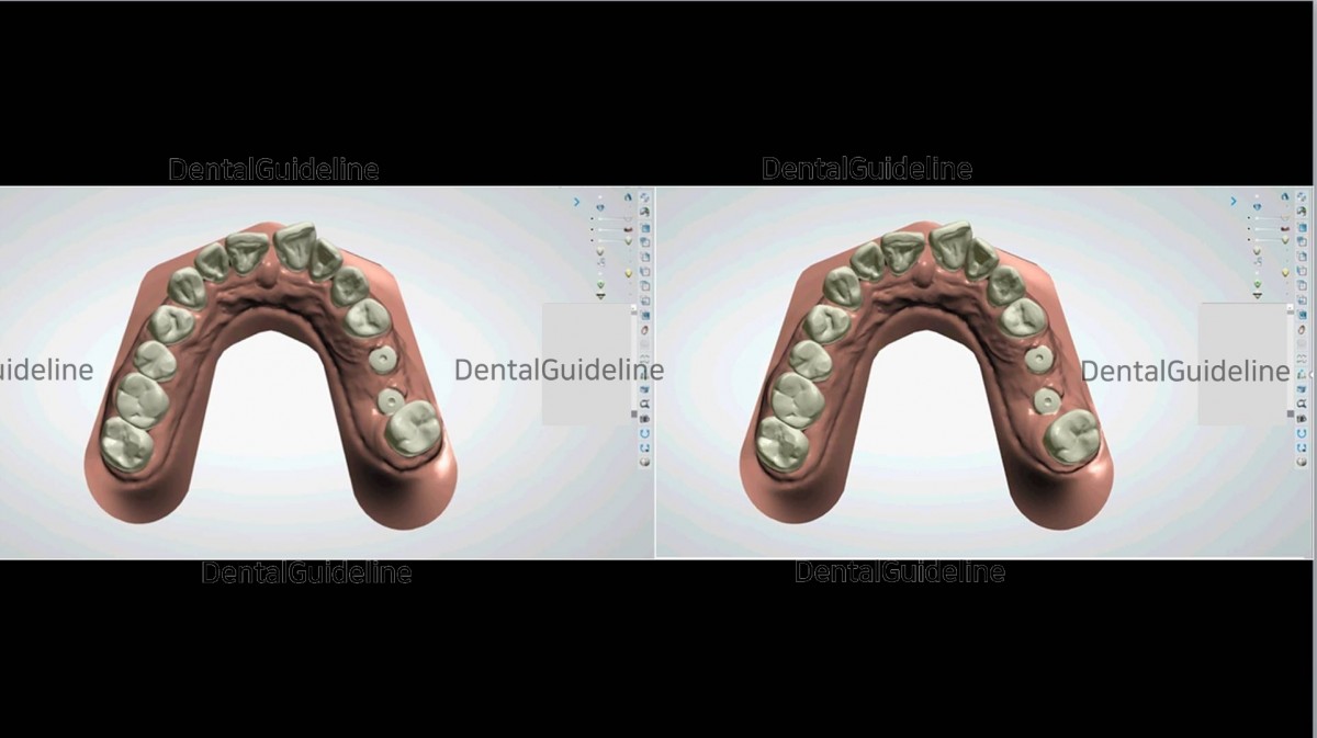
10. 2nd molar movement simulation – before and after
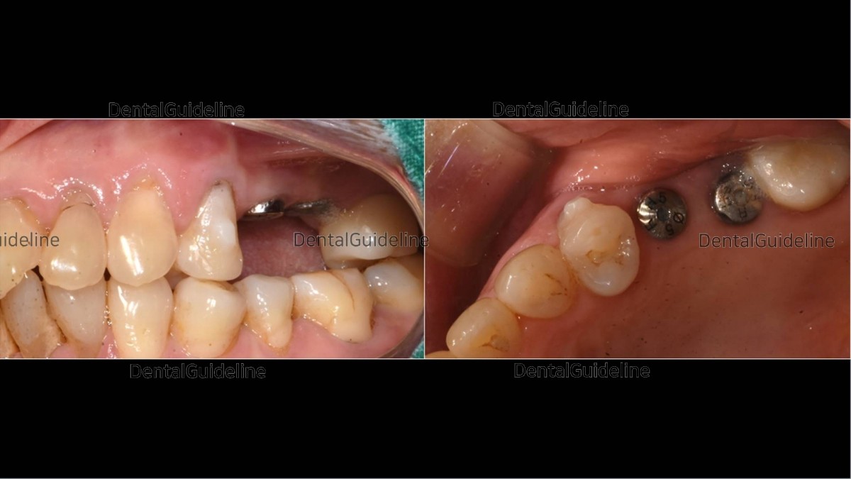
11. Resin attachment was made on the surface of 1st premolar to prevent unexpected tooth movement. . The patient was instructed to change the new appliance every 10 days.
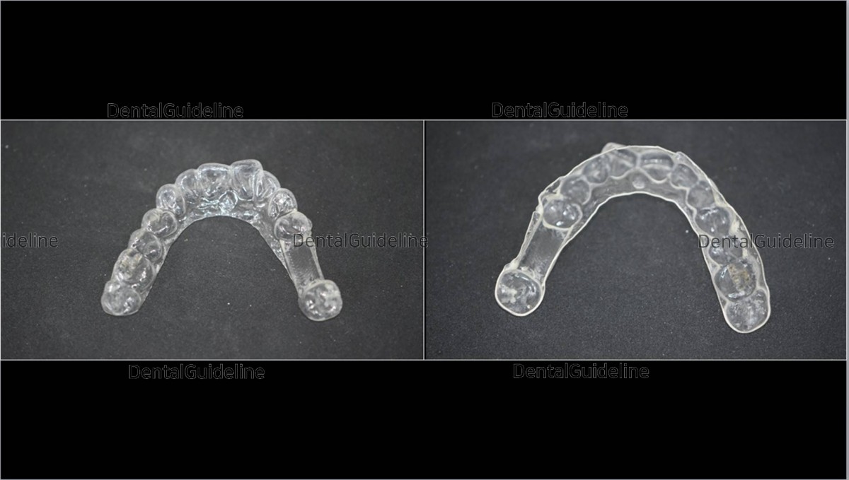
12. Tooth repositioning appliance (one of the series)
.
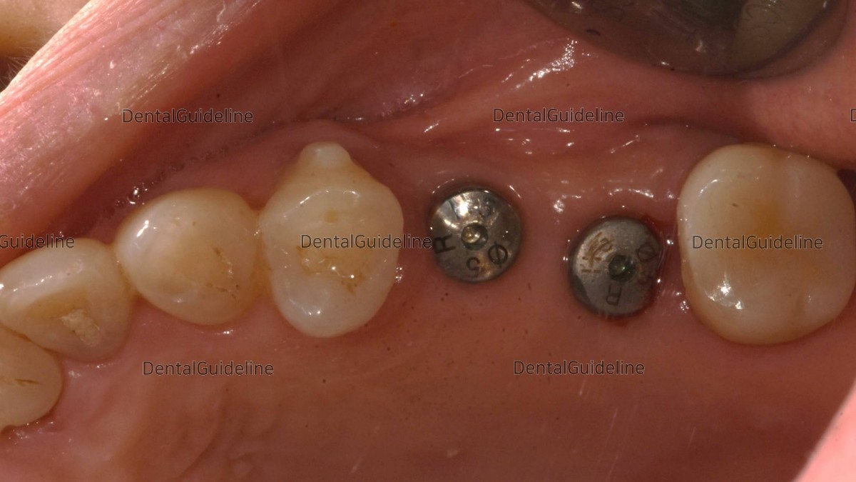
13. After 2.5months of appliance wearing. an intra-oral photo was taken on the day of the ISQ measurement.
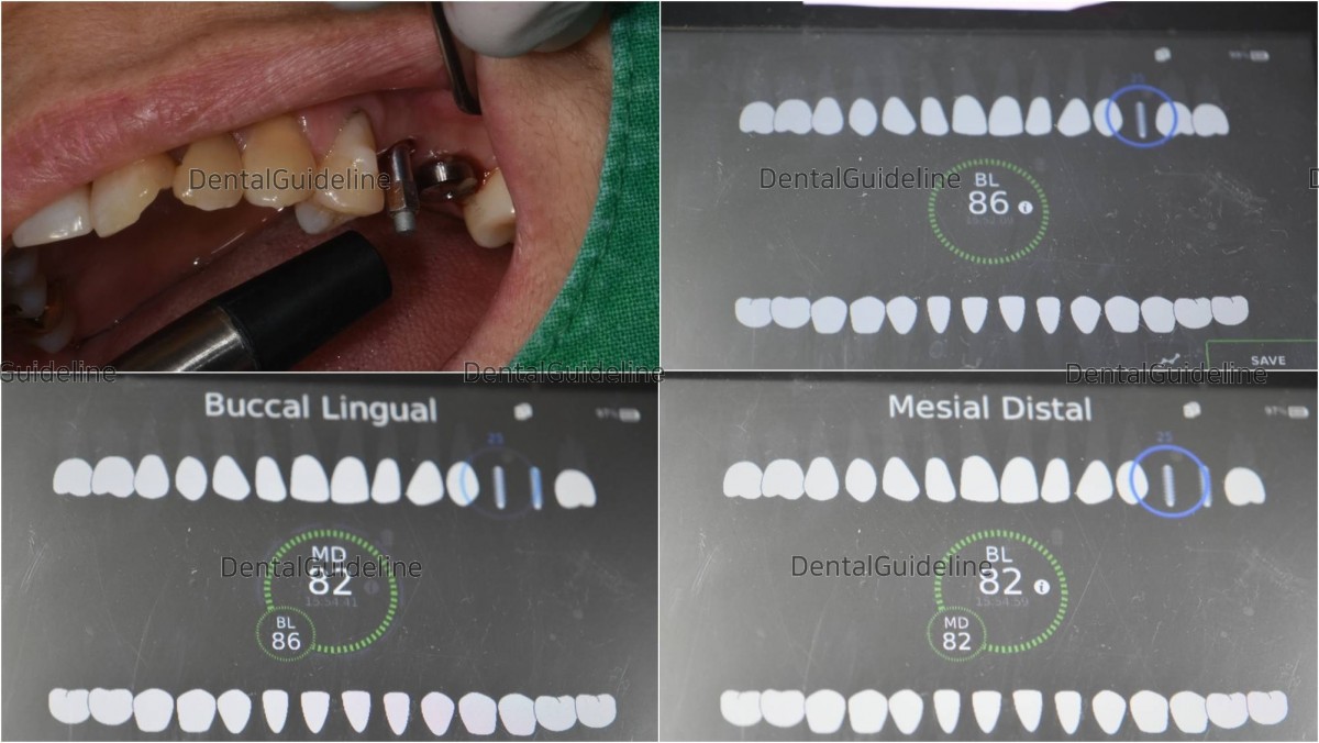
14. ISQ value at the second premolar zone.
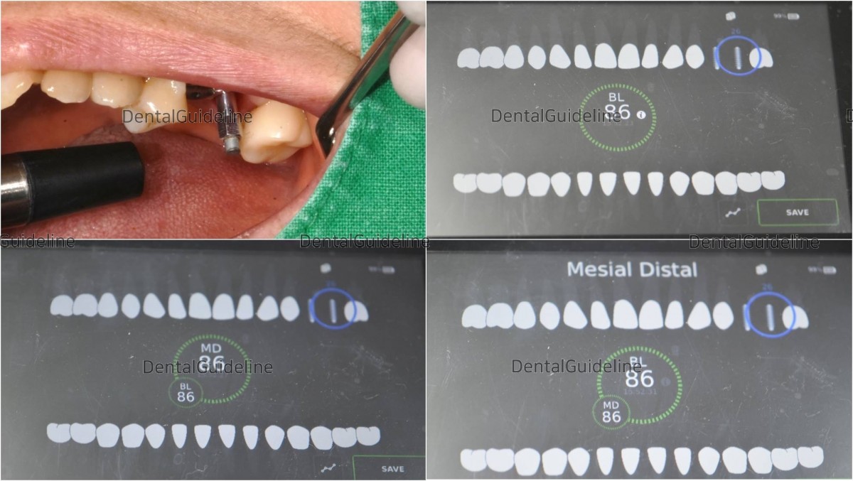
15. ISQ value at the first molar zone.
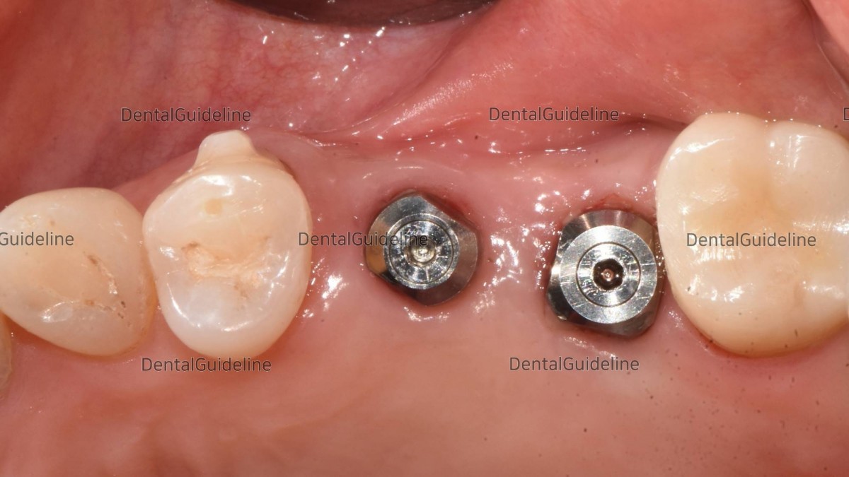
16. Scan-abutments were connected for digital impressions.
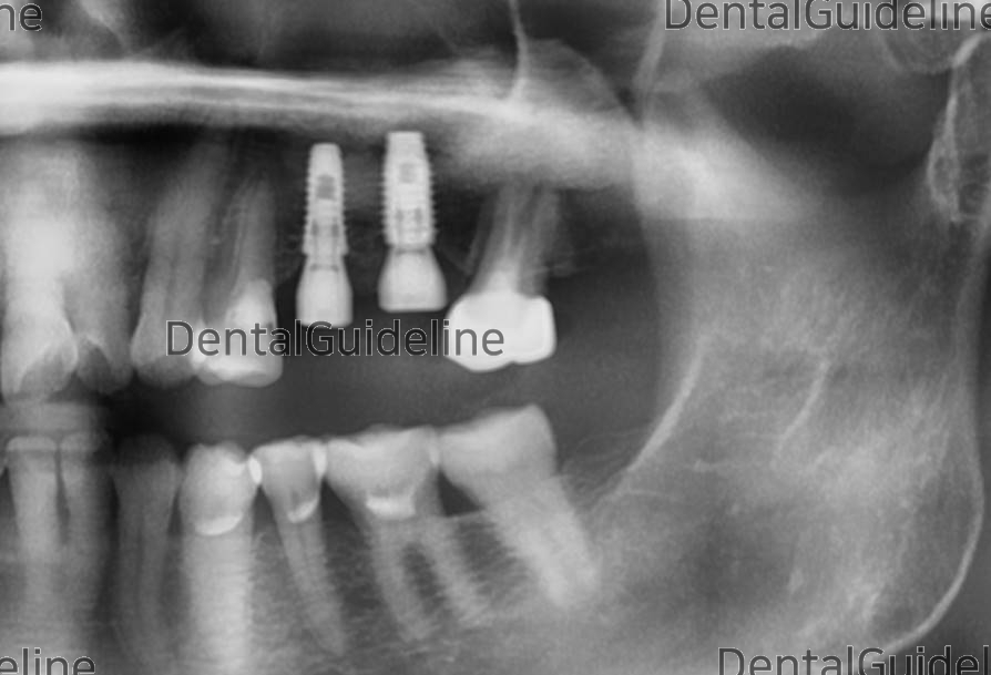
17. A panoramic radiograph was taken to check the secure connection of scan abutments.
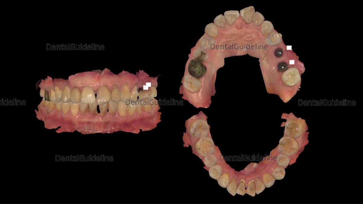
18. . Intra-oral scanning to fabricate custom abutments and provisional resin crowns.
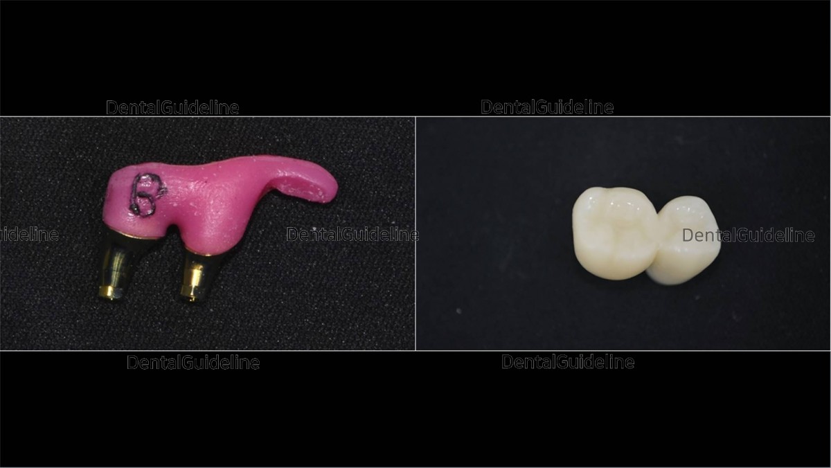
19. The custom abutment, orientation jig, and provisional crown.
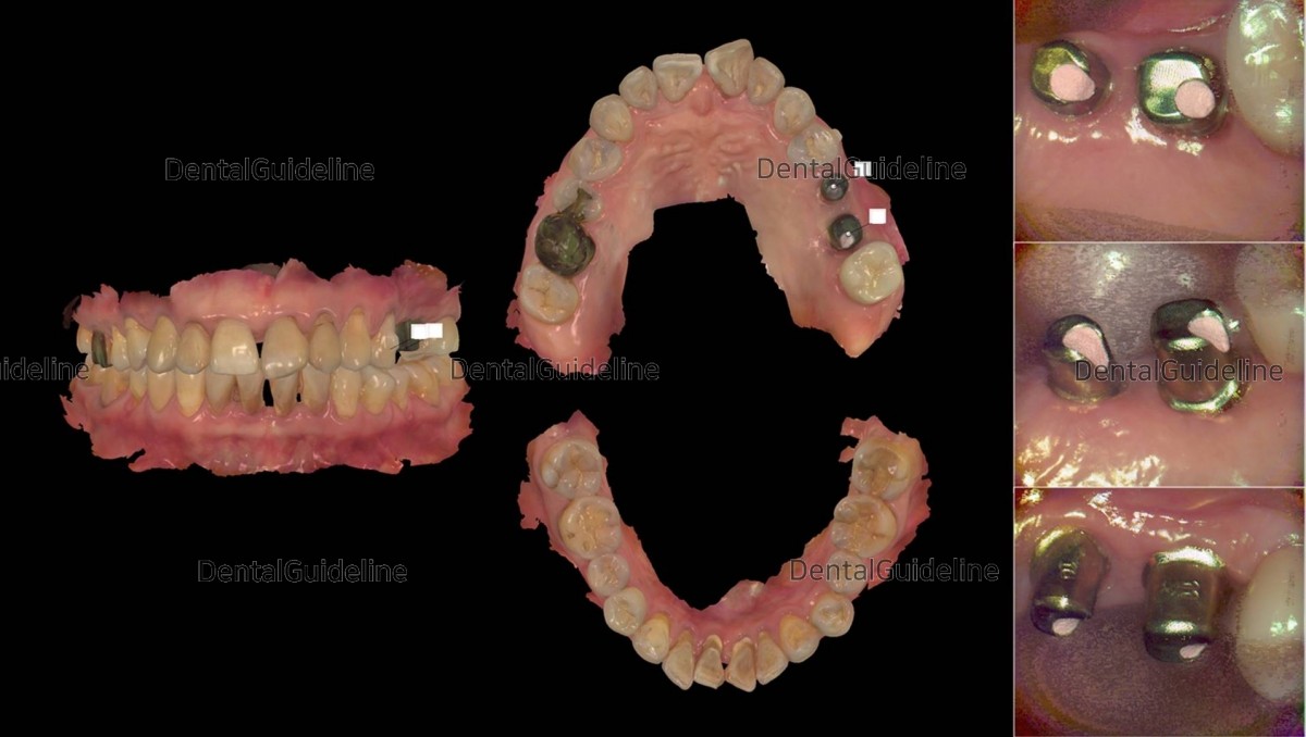
20. Digital impression (intra-oral scanning) for the final restoration.
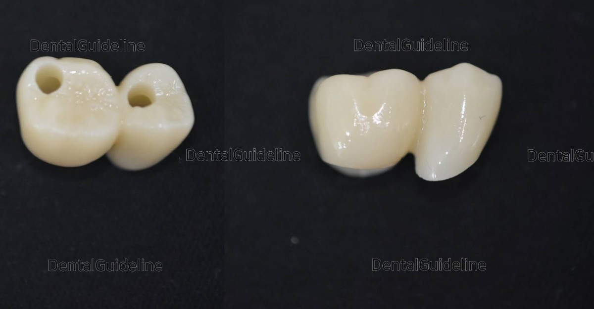
21. The final prosthesis (zirconia)
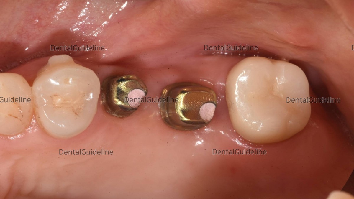
22. provisional crown was removed.
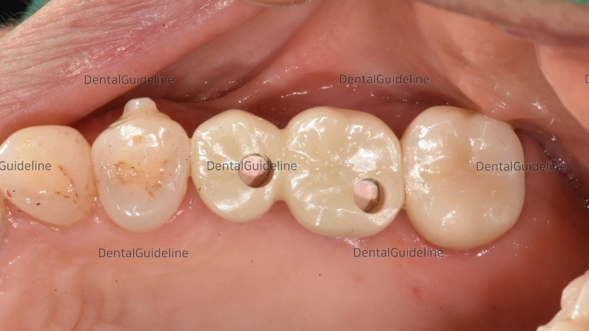 23. Seating trial
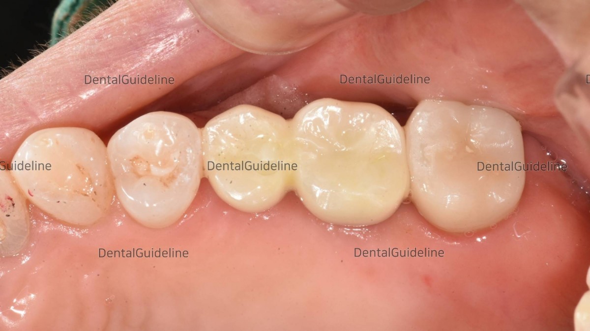
24. cementation and access hole filling.
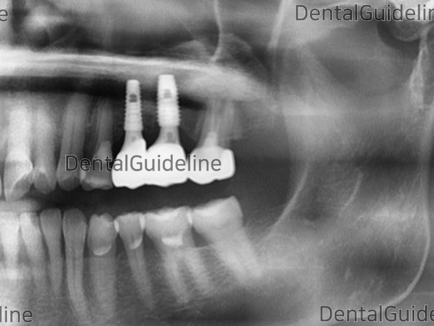
25. Panoramic radiograph after restoration.
0
- PrevMaxillary sinus graft, Tooth uprighting for space regaining (MTM)Nov, 14, 2022
- NextSingle implant installation (video of fixture unpacking) Nov, 14, 2022
There are no registered comment.


