Implantology
Dental Restoration
Periodontics
Dental Labrotary
Two Implants placement with GBR (ARUM DENTISTRY Co.)

HAPPYTOGETHER
Views : 1,010/ Nov, 13, 2022
Views : 1,010/ Nov, 13, 2022
A 52-year-old female patient had experienced intermittent bad taste and foul odor from the old crown on the lower right side.
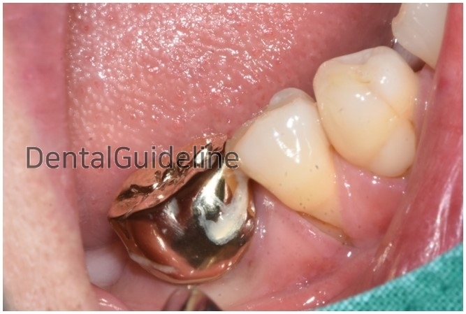
Initial photo
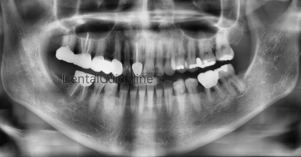
Initial panoramic radiograph.
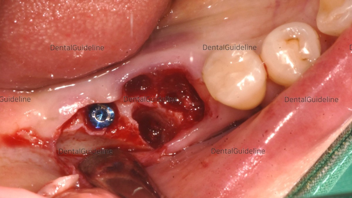
The infected tooth was removed and an implant was placed in the missing area.
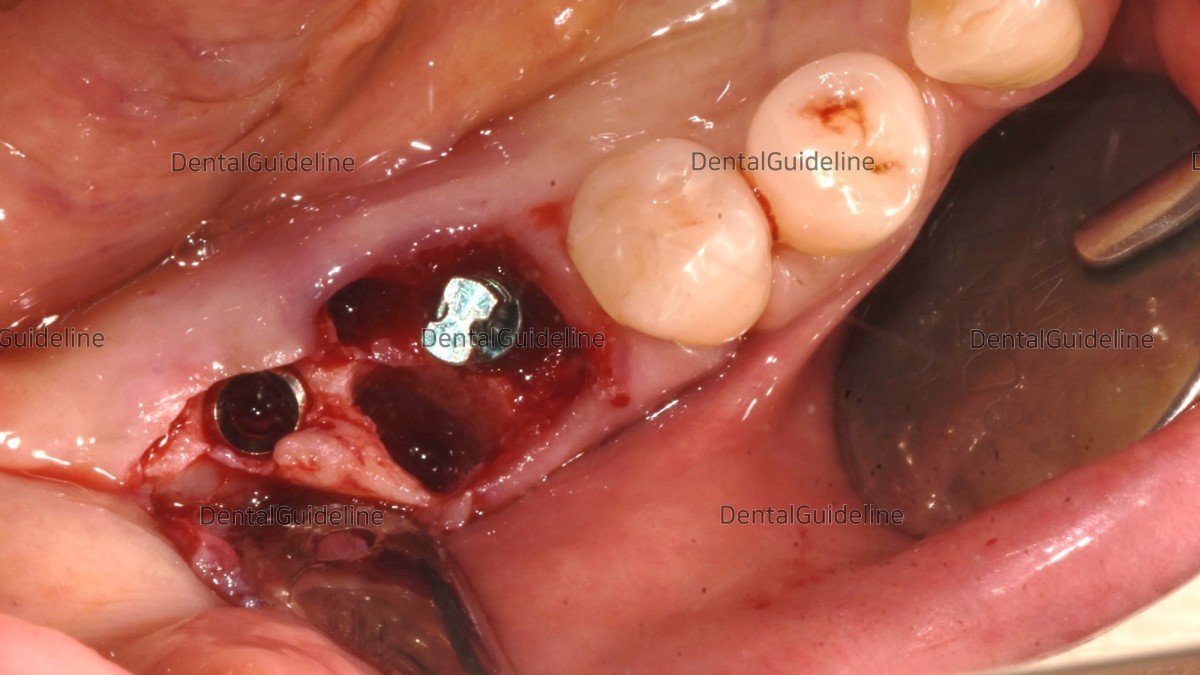
An implant was placed in the extraction socket immediately and a direction pin was screwed into the fixture to verify its position.
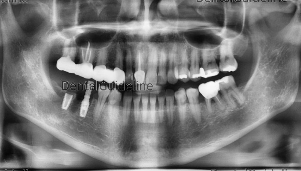
A panoramic radiograph was taken to check the path and relative position of the implants. (ARUM DENTISTRY Co. NB1 Ø4.5/L8.5 X2)

Torque values as initial stability at the 1st and 2nd molar zone.
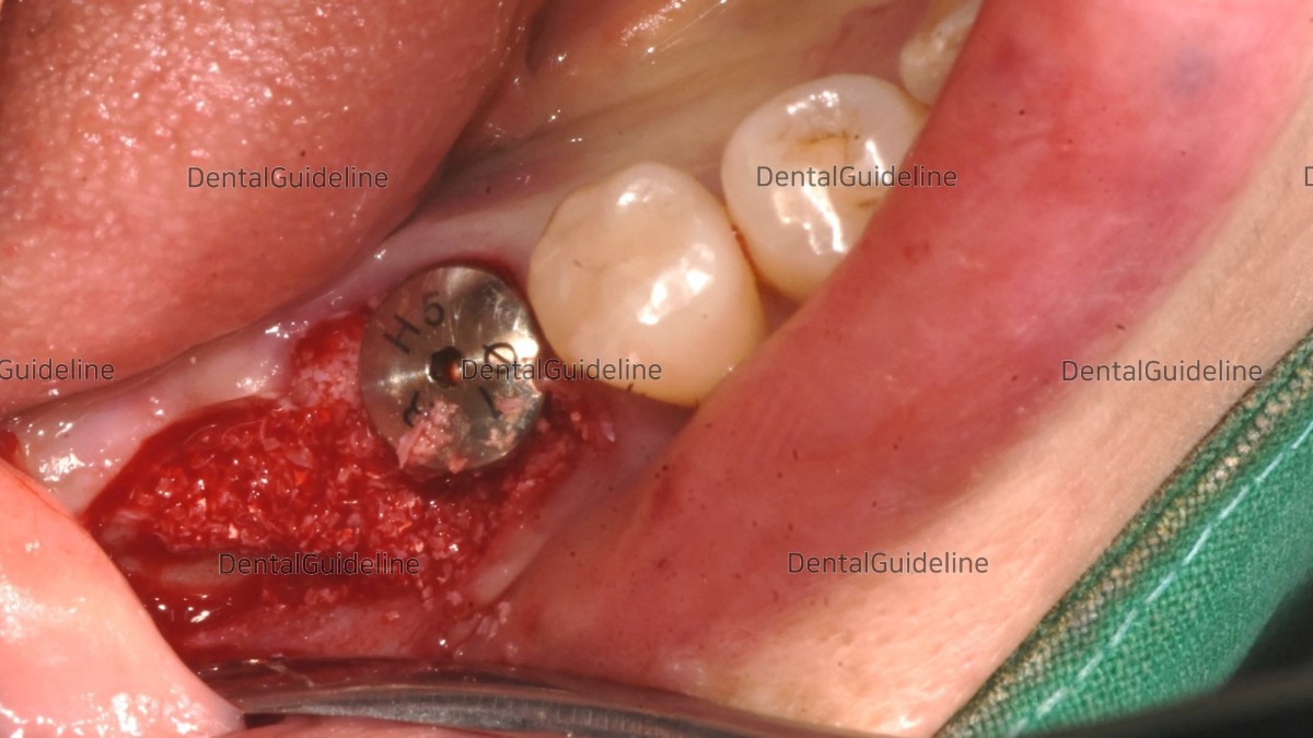
The choice of a healing abutment was engaged in the fixture of the 1st molar zone. And GBR was performed (xenograft).
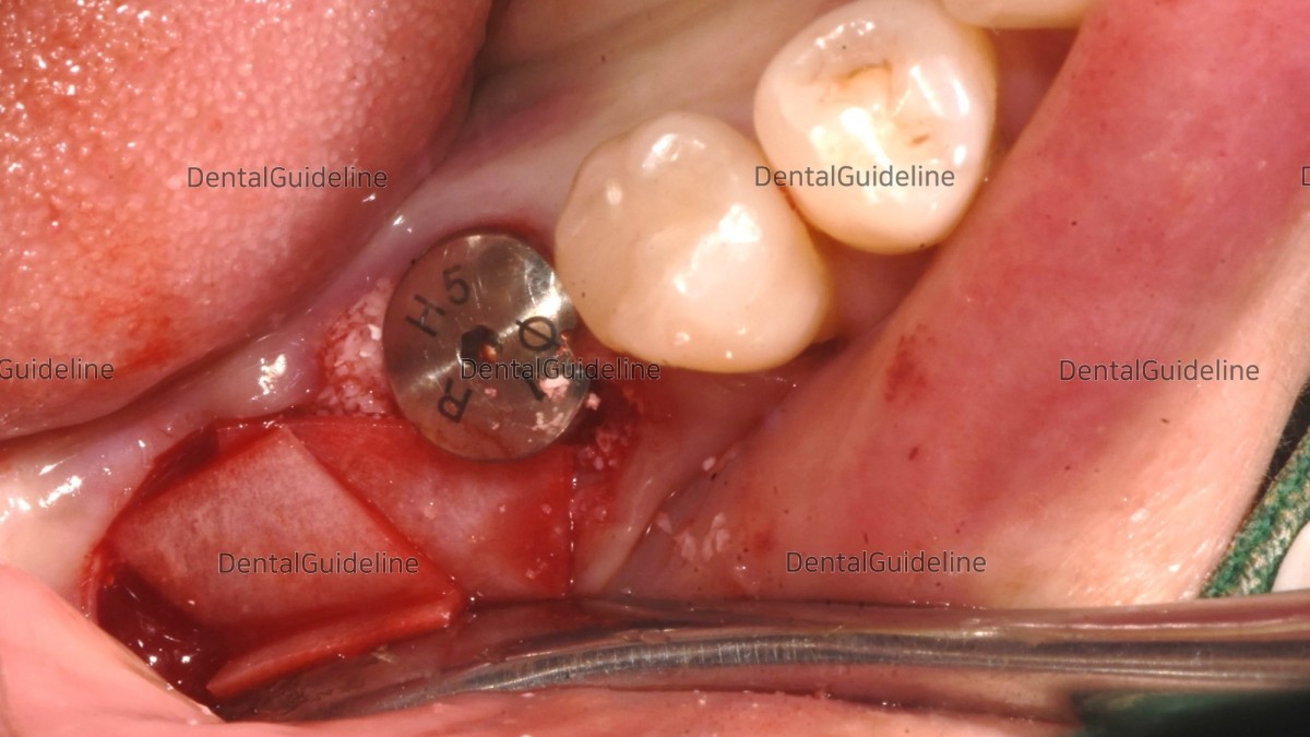
Absorbable collagen membranes were applied.
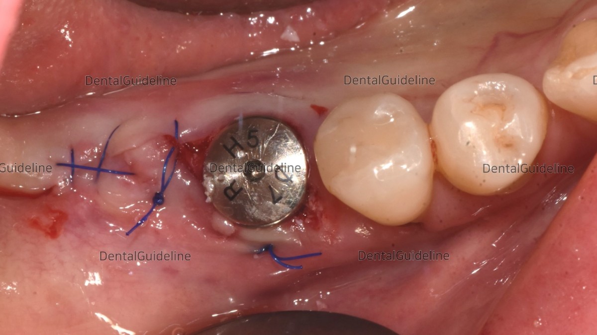
The flap was sutured close. (suture material-nylon)
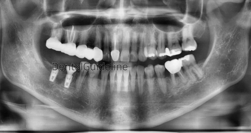
Panoramic radiograph after the surgery.
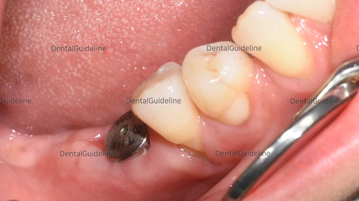
4 weeks post-op.
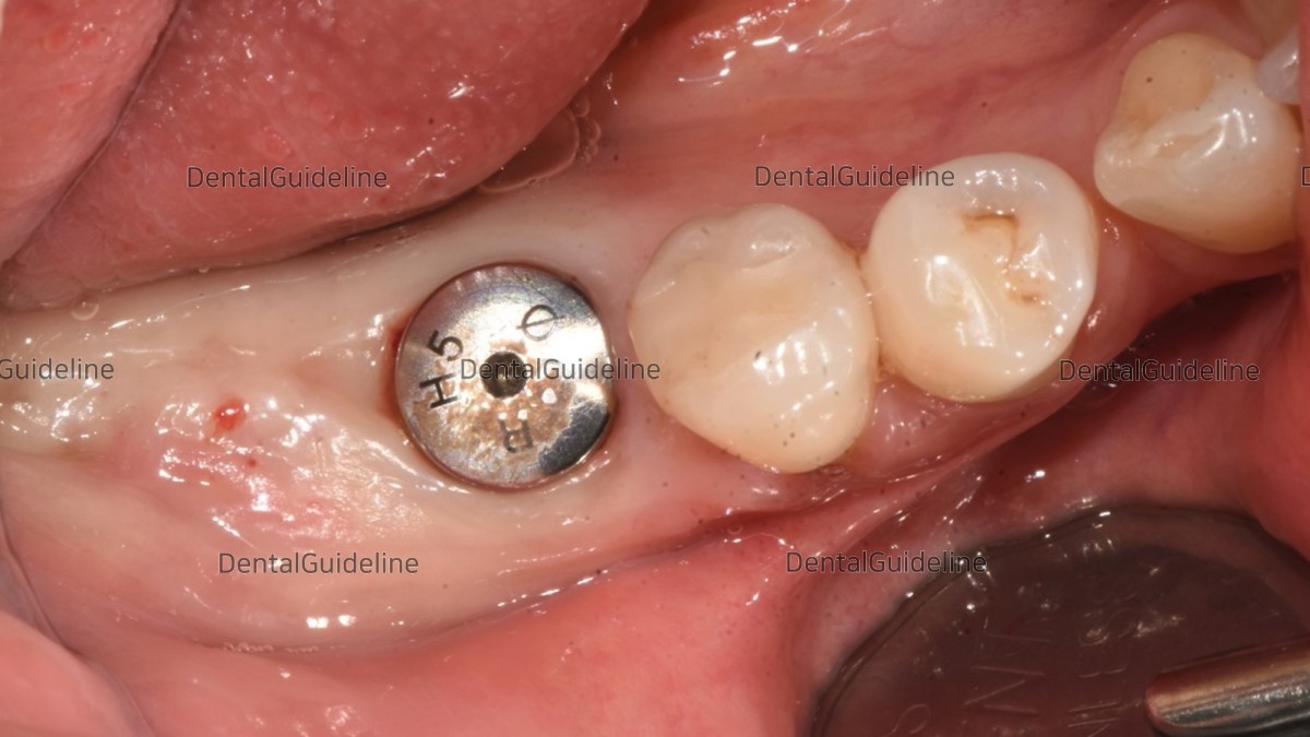
9weeks post-op. The anesthetic shot was given for implant uncovery.
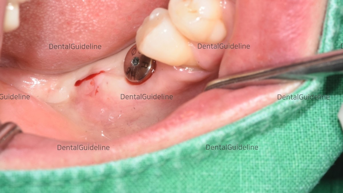
An incision was given on the inner margin of the future position of the healing abutment in this case of insufficient attached gingiva.
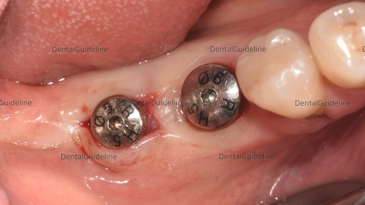
A smaller-sized healing abutment was engaged first.
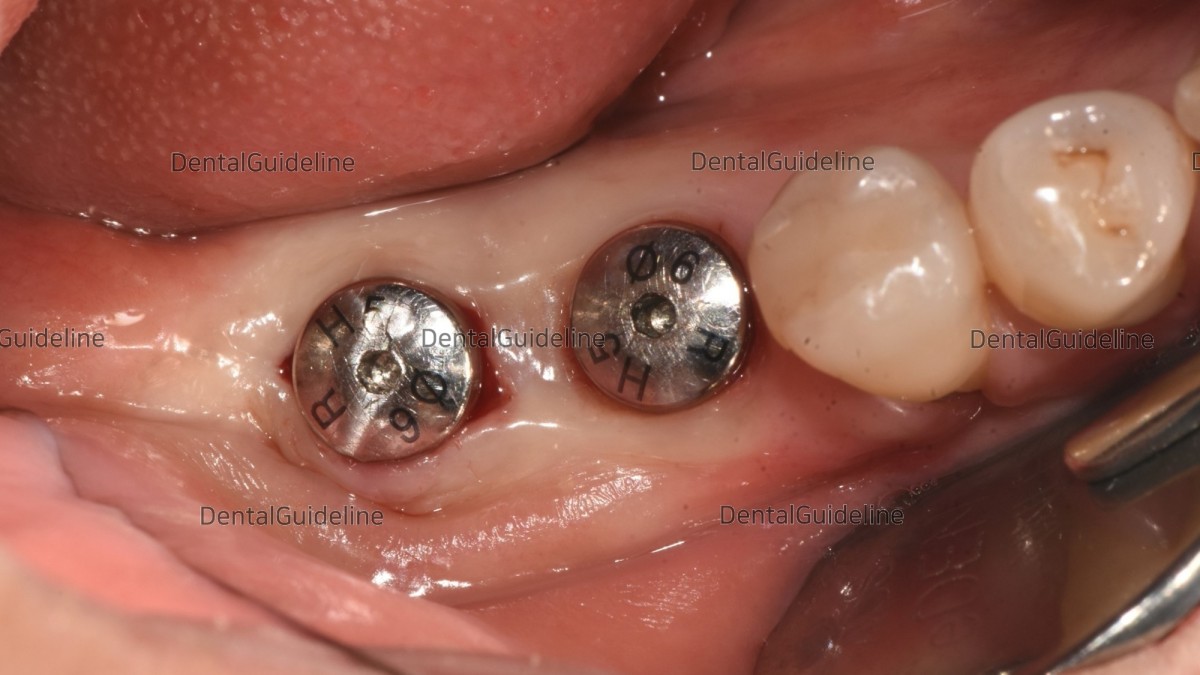
The final-sized healing abutment was engaged.
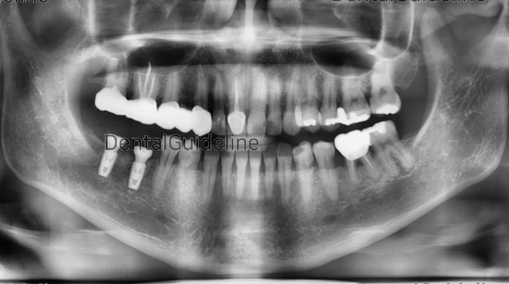
A panoramic radiograph was taken after the implant uncovery.
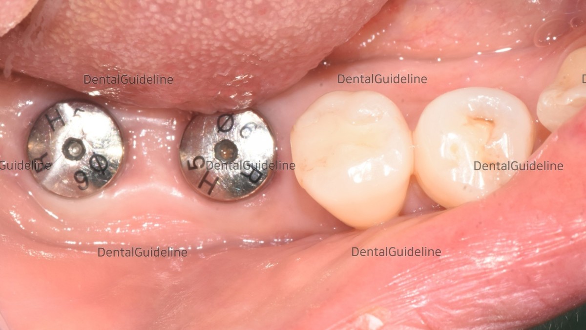
An intraoral photo was taken on the day of impression taking.
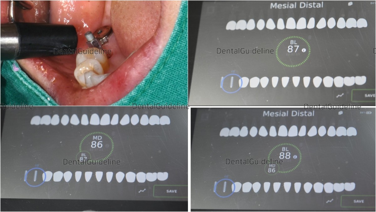
ISQ reading at 1st molar zone.
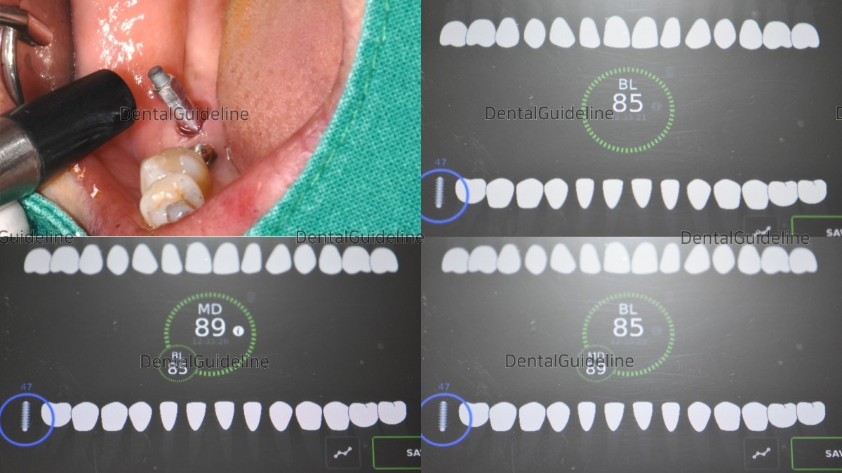 ISQ reading at 2nd molar zone. And impression taking was performed on the day of the ISQ check.

Customized abutments and prostheses.
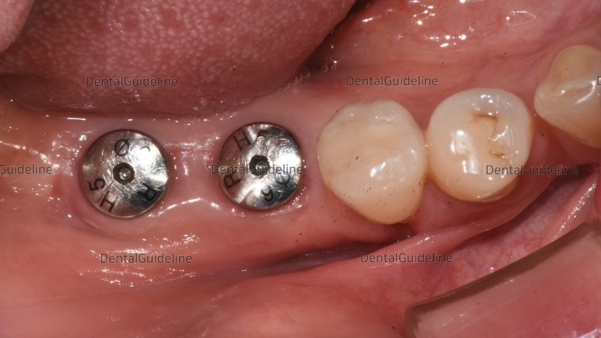
Intraoral view on the day of implant restoration

The seating jig seemed helpful for the connection of the abutment even in the Hexa structure.
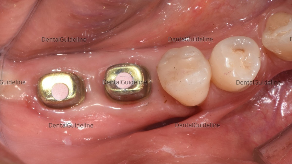
Screw hole filling
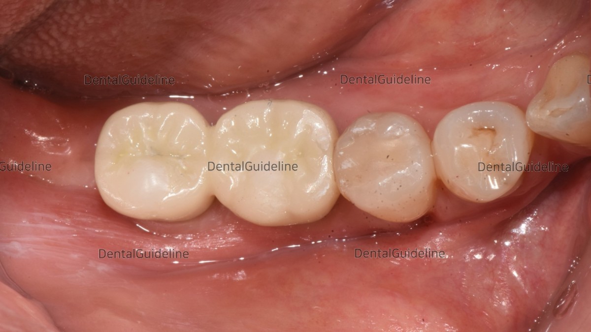
Cementation and hole filling
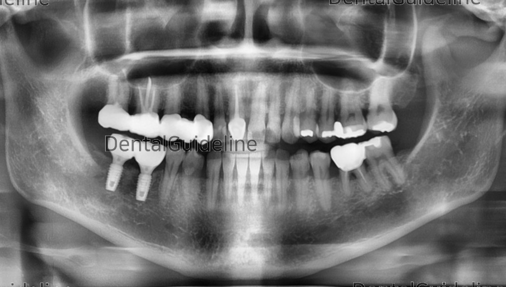
Panoramic view after crown cementation.
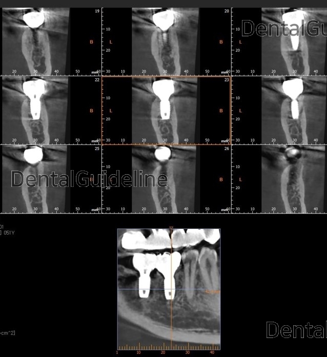
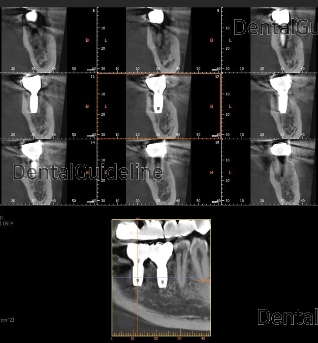
CBCT

Initial photo

Initial panoramic radiograph.

The infected tooth was removed and an implant was placed in the missing area.

An implant was placed in the extraction socket immediately and a direction pin was screwed into the fixture to verify its position.

A panoramic radiograph was taken to check the path and relative position of the implants. (ARUM DENTISTRY Co. NB1 Ø4.5/L8.5 X2)

Torque values as initial stability at the 1st and 2nd molar zone.

The choice of a healing abutment was engaged in the fixture of the 1st molar zone. And GBR was performed (xenograft).

Absorbable collagen membranes were applied.

The flap was sutured close. (suture material-nylon)

Panoramic radiograph after the surgery.

4 weeks post-op.

9weeks post-op. The anesthetic shot was given for implant uncovery.

An incision was given on the inner margin of the future position of the healing abutment in this case of insufficient attached gingiva.

A smaller-sized healing abutment was engaged first.

The final-sized healing abutment was engaged.

A panoramic radiograph was taken after the implant uncovery.

An intraoral photo was taken on the day of impression taking.

ISQ reading at 1st molar zone.
 ISQ reading at 2nd molar zone. And impression taking was performed on the day of the ISQ check.
Customized abutments and prostheses.

Intraoral view on the day of implant restoration

The seating jig seemed helpful for the connection of the abutment even in the Hexa structure.

Screw hole filling

Cementation and hole filling

Panoramic view after crown cementation.


CBCT
0
- PrevSingle implant installation (video of fixture unpacking)Nov, 13, 2022
- NextApical Surgery in the esthetic zone and Restorations. SURGYBONE. Nov, 13, 2022
There are no registered comment.


