Implantology
Dental Restoration
Dental Labrotary
Implant in the molar zone of both jaws

HAPPYTOGETHER
Views : 3,715/ Jan, 29, 2023
Views : 3,715/ Jan, 29, 2023
<GCljoo>
A 55-year-old female patient had
bilateral problems in both jaws.
It was decided to proceed with implant-supported restoration in the left molar part first.
She had been taking hypertension medication for a long time.
 ▲first visit photo
 ▲Panoramic radiograph before the implant surgery in the lower left area.
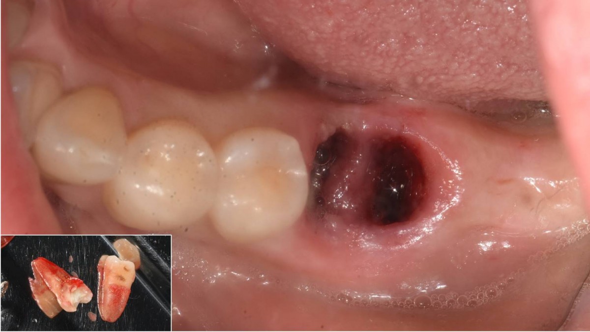 ▲Extraction.
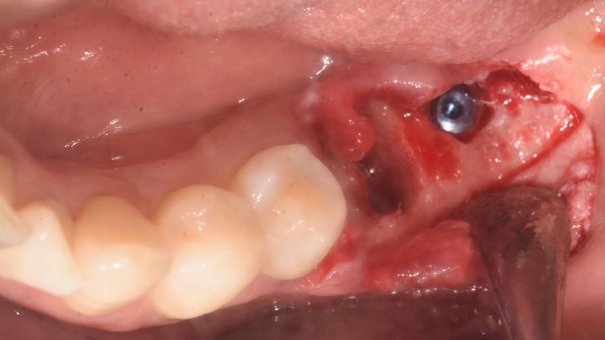 ▲implant placement in the edentulous area. Arum Dentistry NB1 5*8.5 (10Ncm).
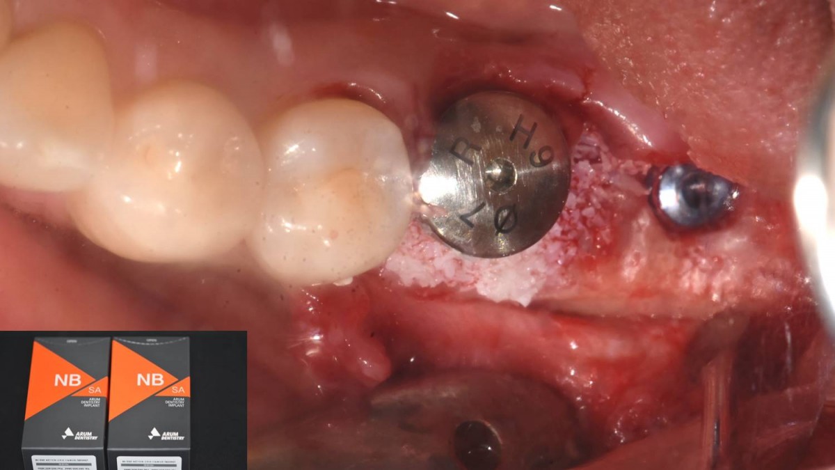 ▲Immediate implant placement, healing abutment engagement. Arum Dentistry NB1 5*10 (30Ncm).
 ▲GBR
▲GBR ▲Collagen membrane
  ▲Suture.
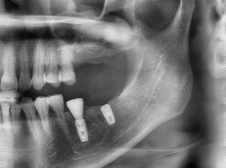 ▲post-op panoramic radiograph.
 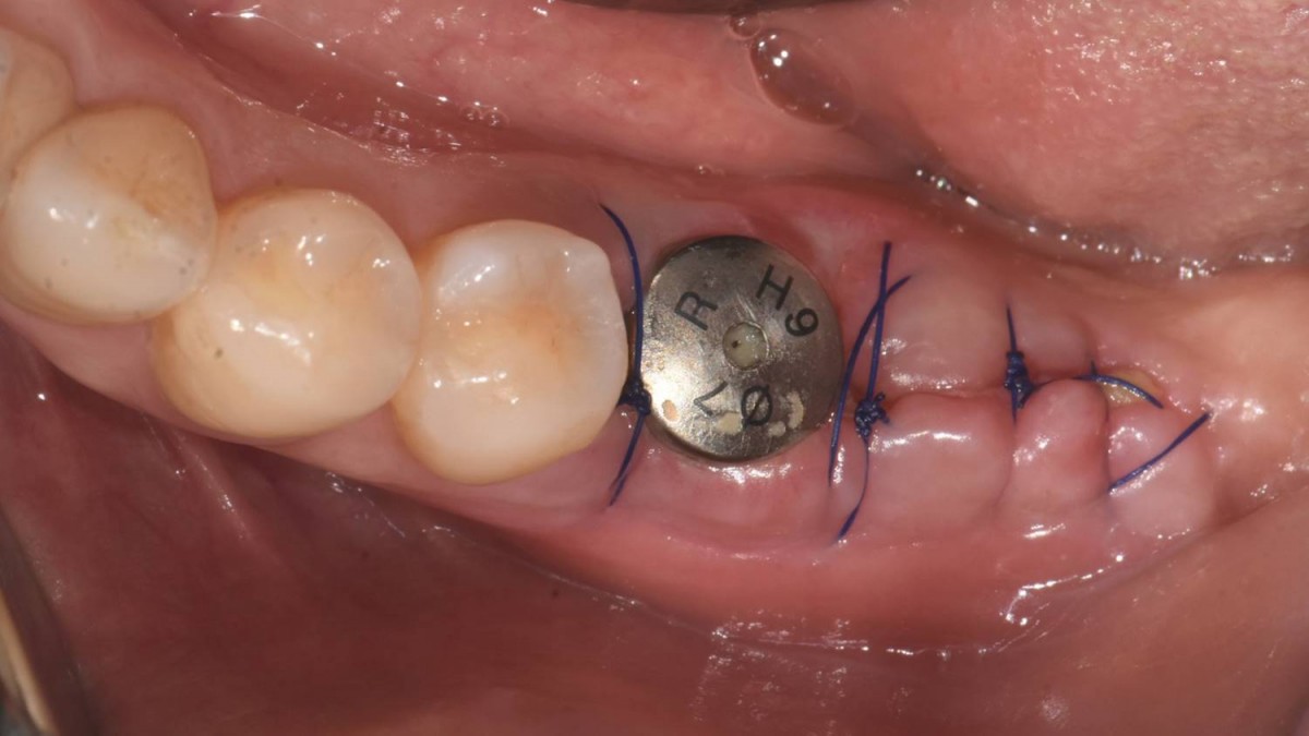 ▲1 week post-op.
 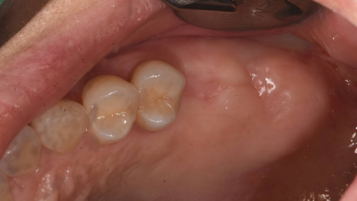 ▲Upper jaw. Intraoral photo before implant surgery.
 ▲A maxillary sinus graft was performed 3 months ago.
 ▲Arum Dentistry NB1 5*11.5 (30Ncm) at the first molar zone.
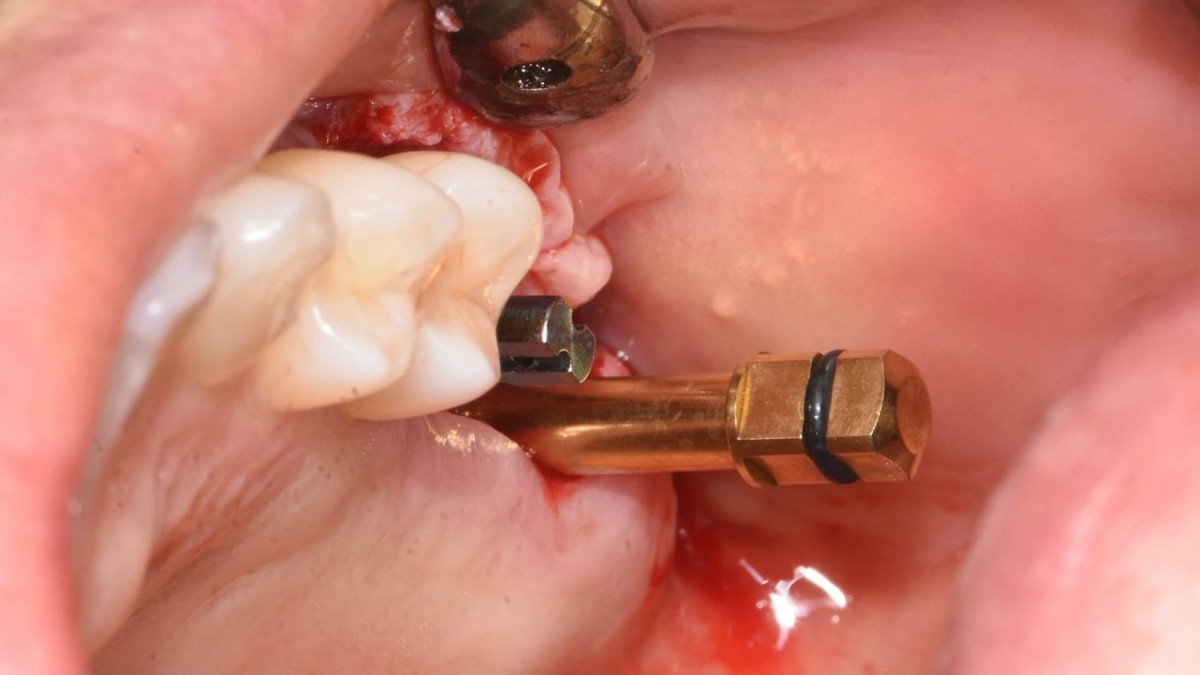 ▲Check the orientation of the two implants using the direction pin.
 ▲Arum Dentistry NB1 5*11.5 (15Ncm) at the second molar zone
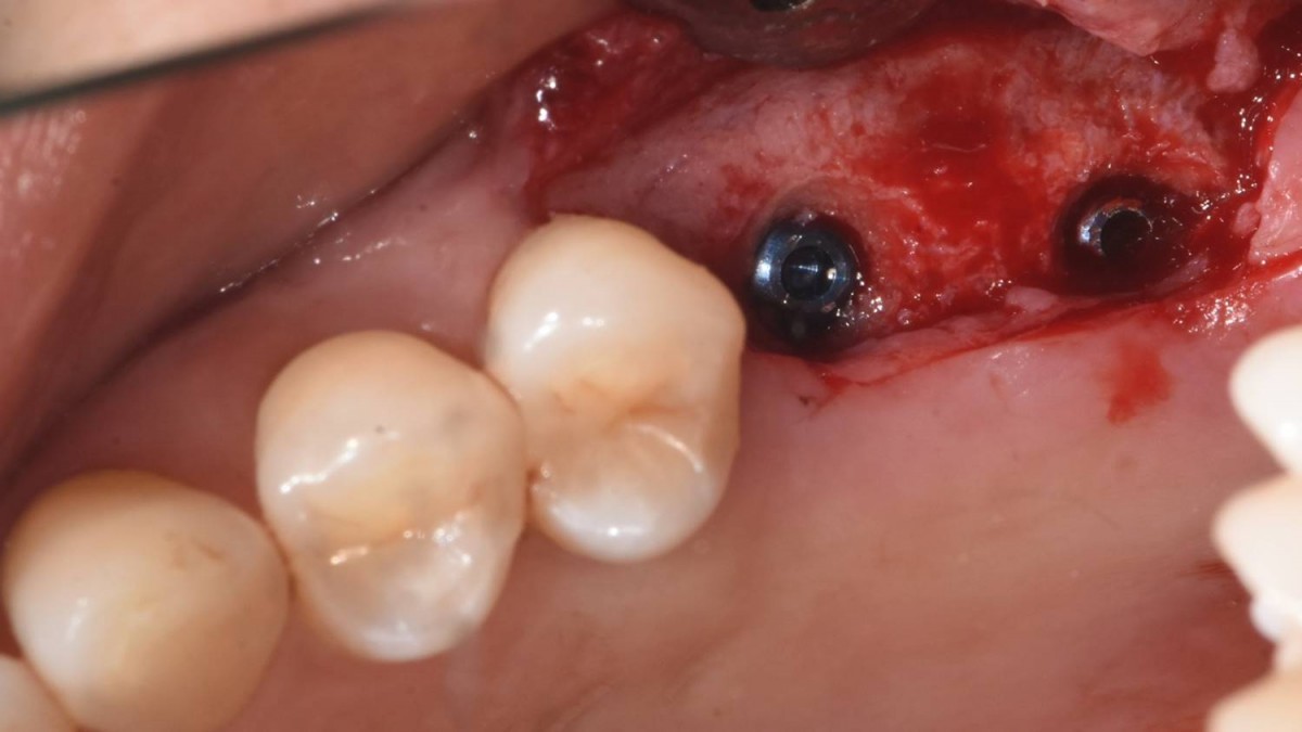 ▲Arum Dentistry NB1 5*11.5 (first molar-30Ncm, second molar-15Ncm).
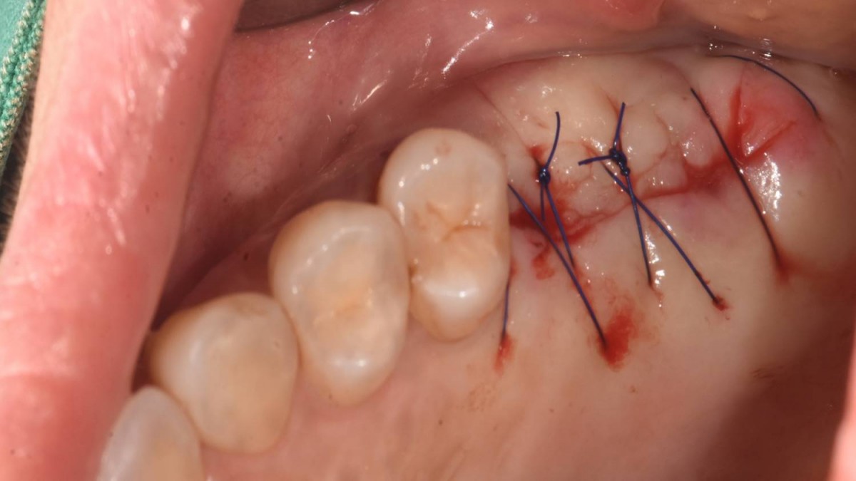 ▲Suture.
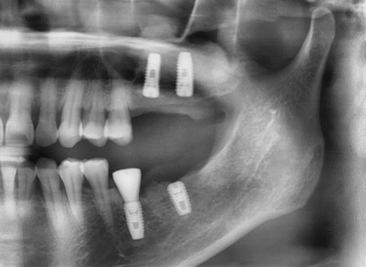 ▲. Panoramic radiograph after implant placement in the maxilla.(3 weeks after implant placement in the mandible)
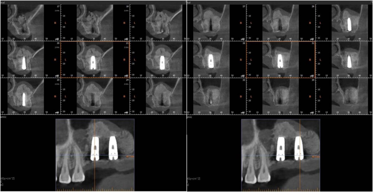 ▲CBCT
▲CBCT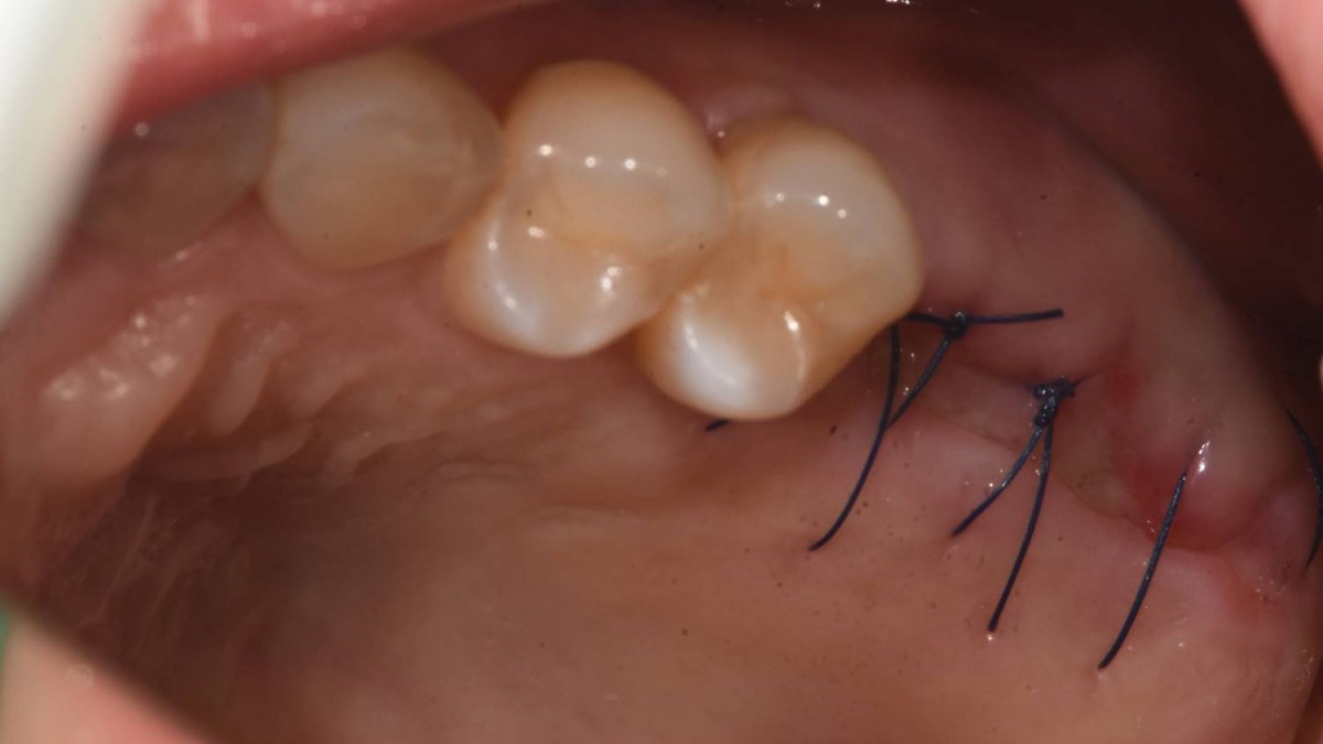 ▲1 week post-op.
 ▲Upper ridge-4 weeks post-op. Lower ridge-7 weeks post-op.
 ▲Healing abutment engagement at the second molar zone. (3 months post-op.)
 ▲ISQ reading at the first molar zone.
 ▲ ISQ reading at the first molar zone.
 ▲Implant uncovery. 4 months post-op.
 ▲Intraoral photo on the day of the ISQ reading in the maxilla.
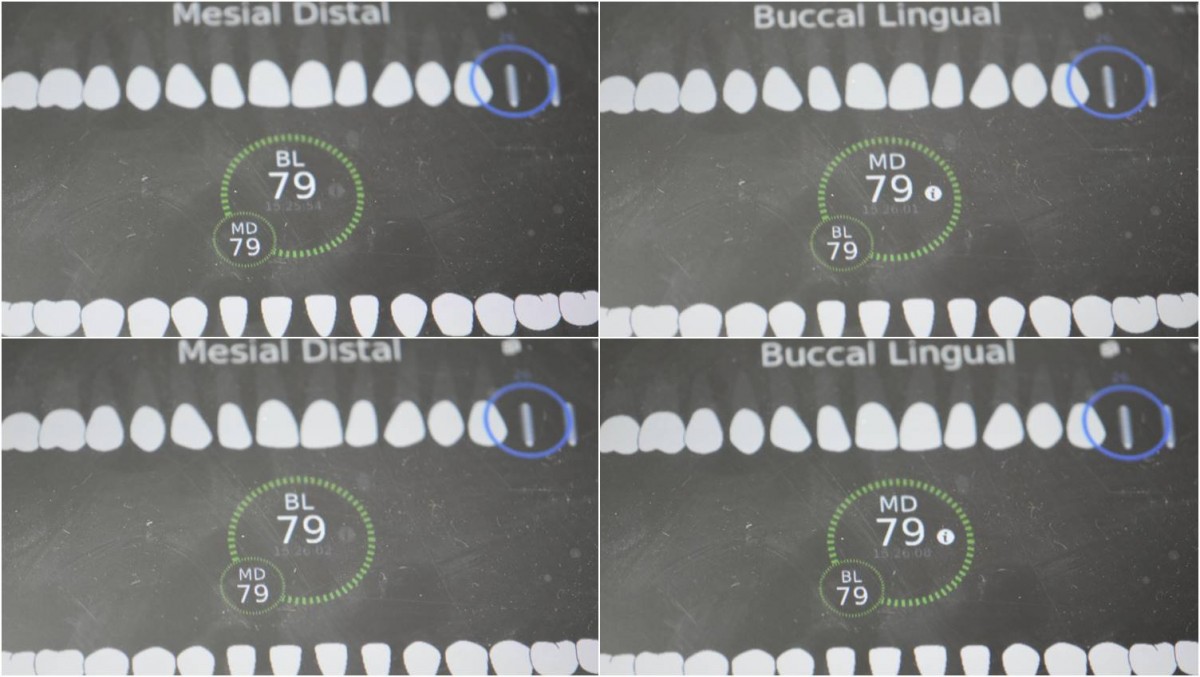 ▲ISQ reading at the first molar zone of the maxilla.
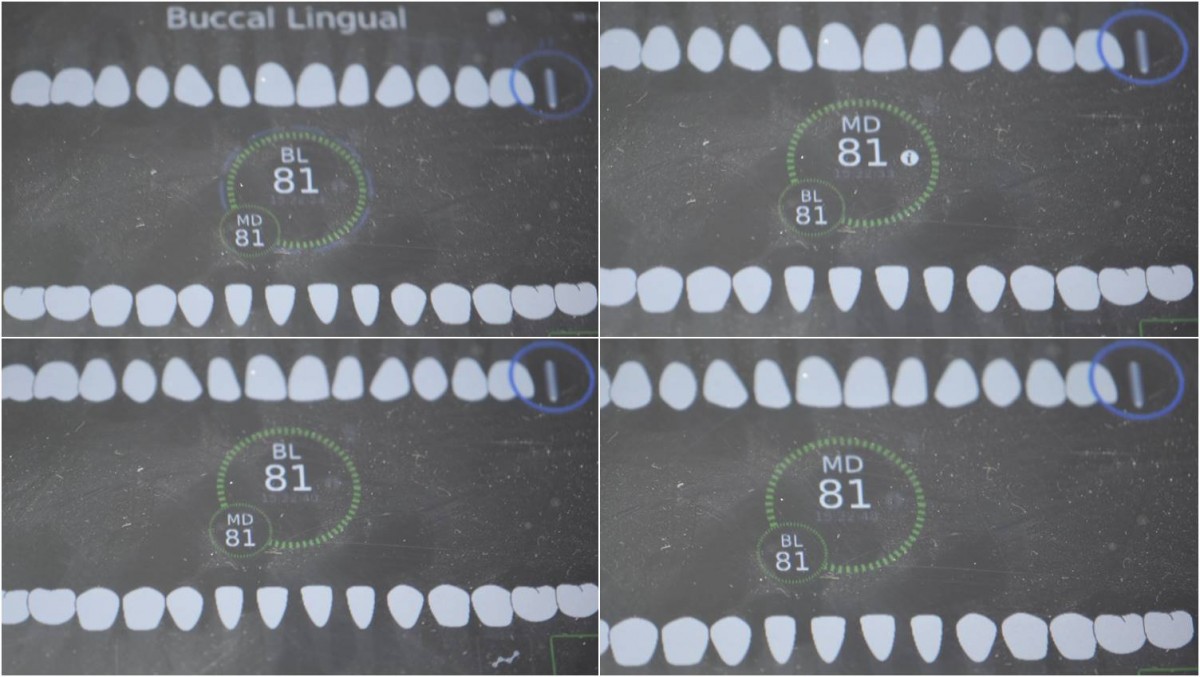 ▲ISQ reading at the second molar zone of the maxilla
 ▲To create a gingival shape of an appropriate size, HA of a different size from the existing one was applied.
 ▲To create a gingival shape of an appropriate size, HA of a different size(diameter and height) from the existing one was applied.
 ▲Custom abutment and crown on the maxillary working model.
 ▲Custom abutment and crown on the mandibular working model.
 ▲Abutment connection.
 ▲Crown seating trial
 ▲ Intraoral photo after cementation.
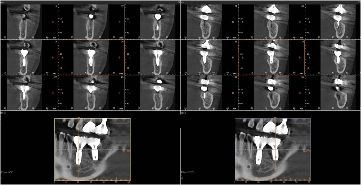 ▲CBCT after cementation of prosthesis
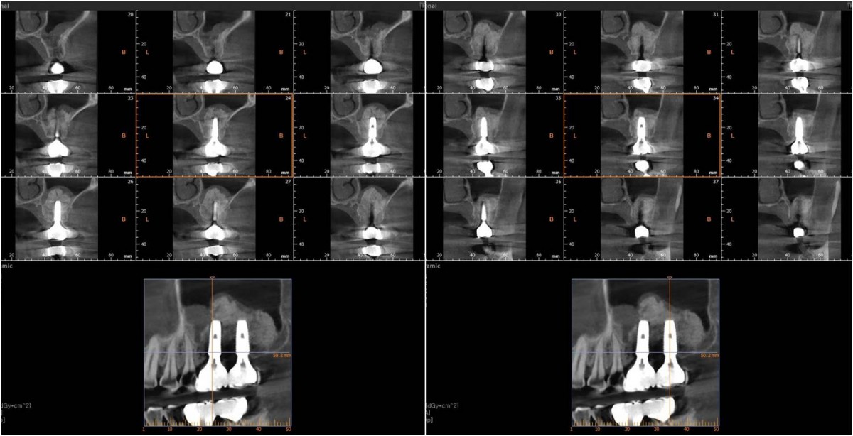 ▲CBCT after cementation of prosthesis
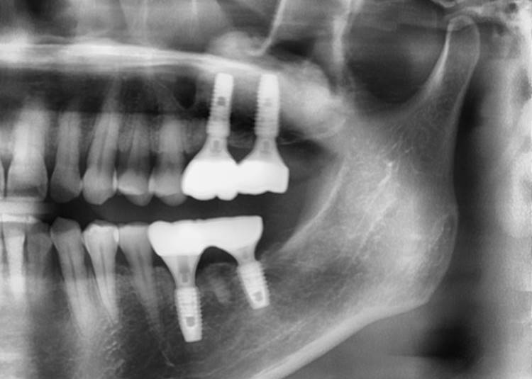 ▲Panoramic radiograph after 1 year of the crown delivery.
 ▲CBCT. 1 year after crown delivery.
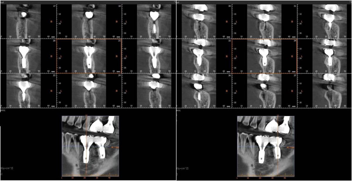 ▲CBCT. 1 year after crown delivery.
0
- PrevVideo, Immediate Implant Placement, Flapless ;PremolarJan, 29, 2023
- NextSingle PFM crown in the esthetic region Jan, 29, 2023
There are no registered comment.


