

Implantology
Dental Restoration
Dental Labrotary
Maxillary Sinus Graft, 2 Implants, Crown Contouring

HAPPYTOGETHER
Views : 622/ Jan, 20, 2023
Views : 622/ Jan, 20, 2023
<GCaks> A 56-year-old male patient had pain-inducing caries, and perio-involved tooth mobility resulted in a tooth fracture at 1st molar. And it was removed months ago. He was a heavy smoker and showed poor oral hygiene.
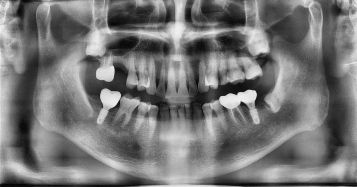 ▲Pre-op panoramic radiograph.
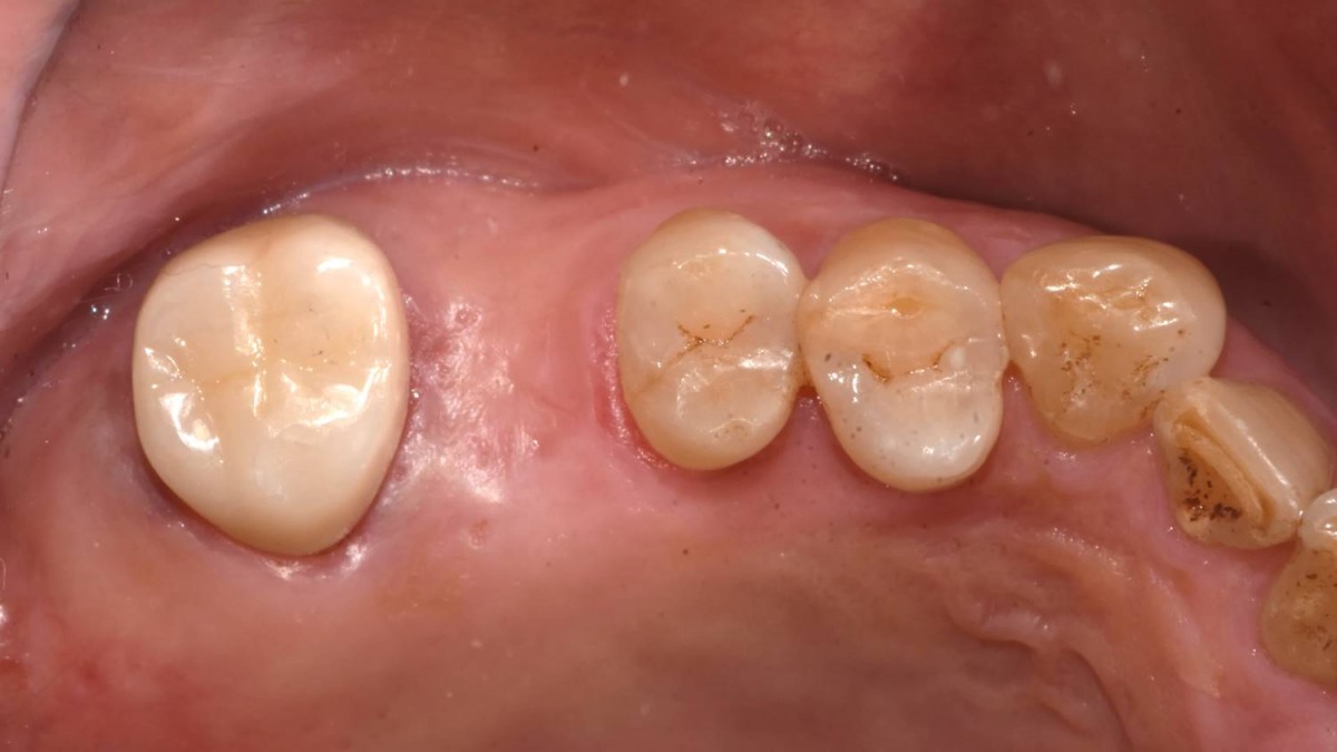 ▲Intraoral view before the maxillary sinus graft
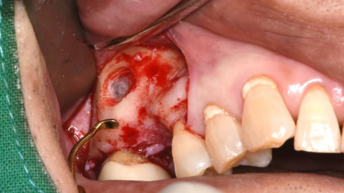 ▲Lateral window using reamer
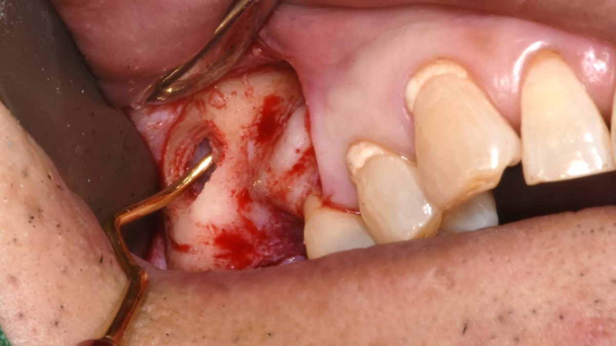 ▲Sinus membrane elevation procedure with all-in-one sinus curette.
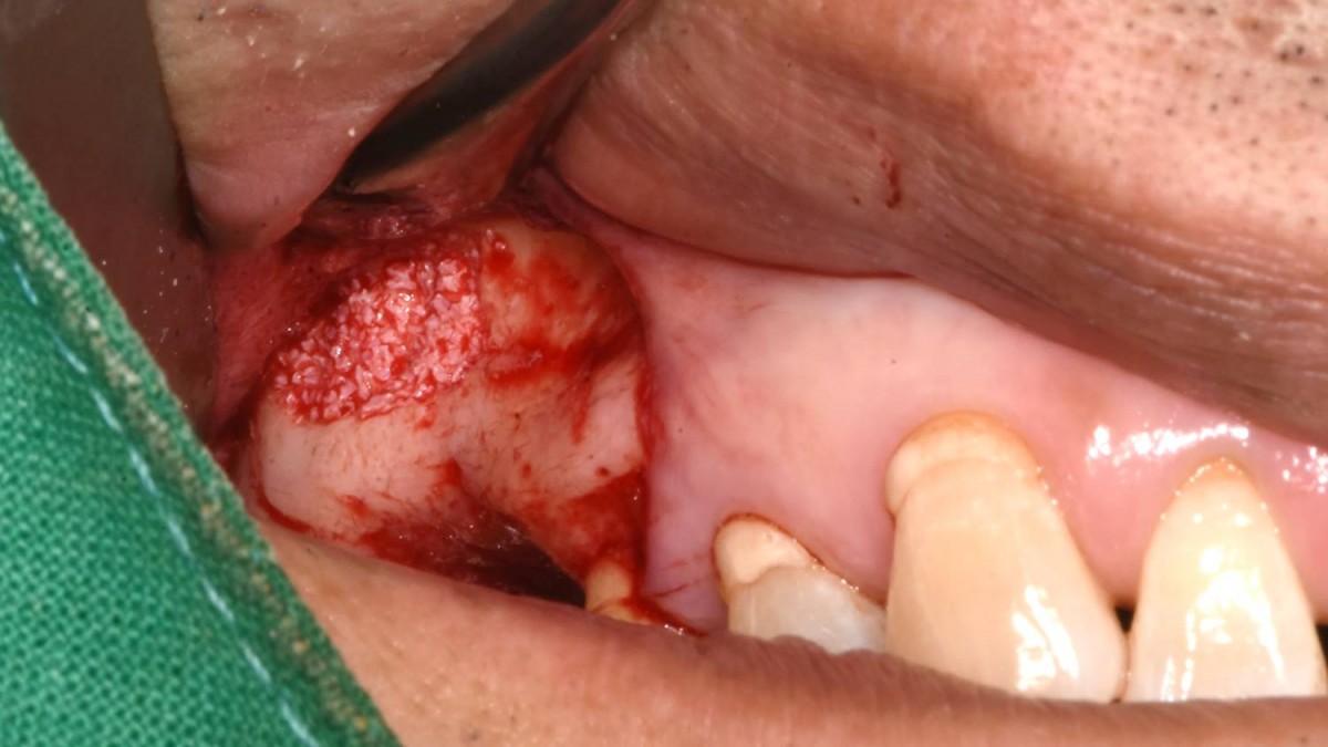 ▲Bone material(Xenograft) was applied into the prepared sinus cavity.
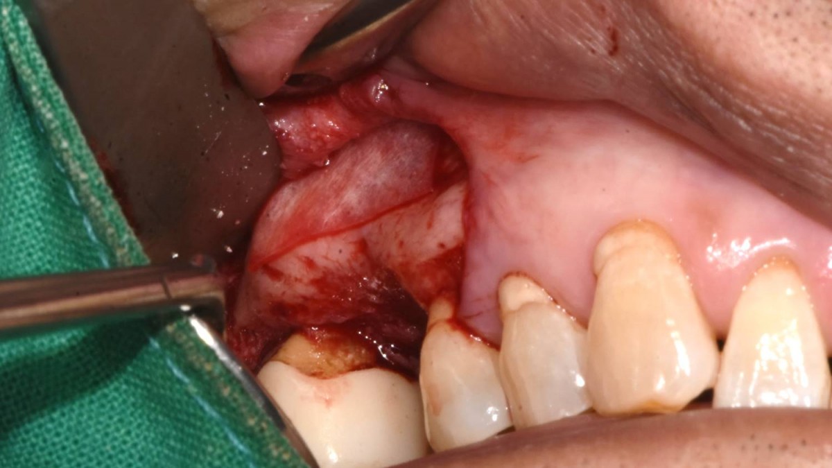 ▲Absorbable collagen membrane was placed on the grafted site. (Lyoplant®)
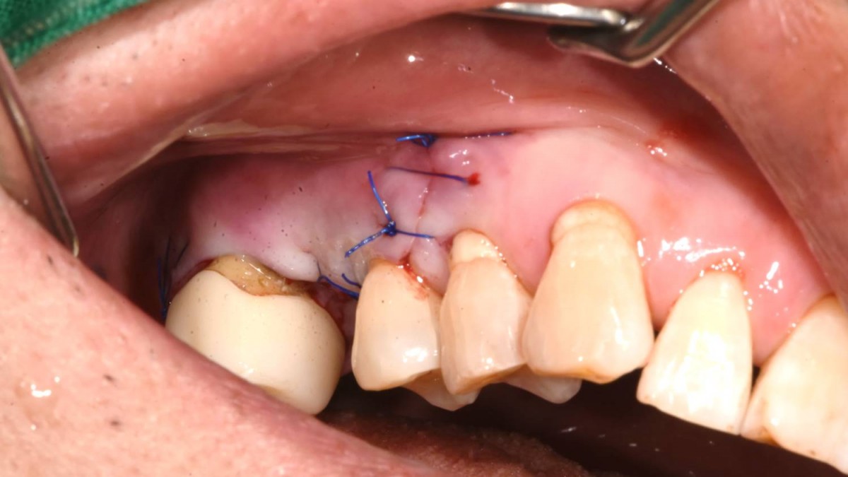 ▲Suture was done (Nylon 4-0)
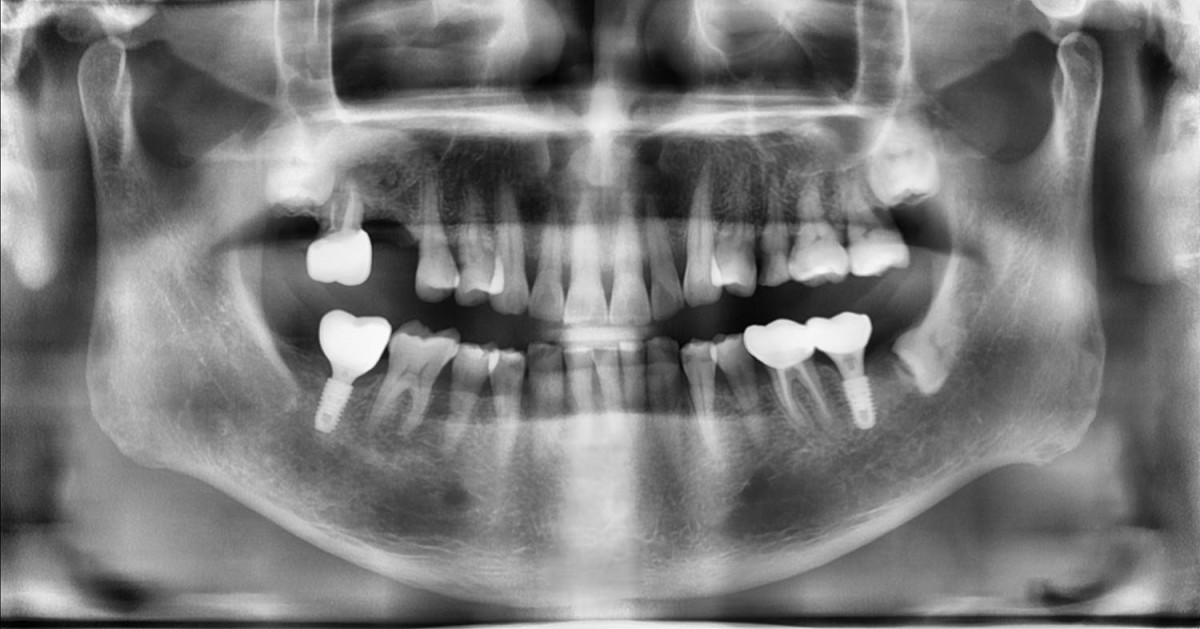 ▲Radiograph was taken right after maxillary sinus graft
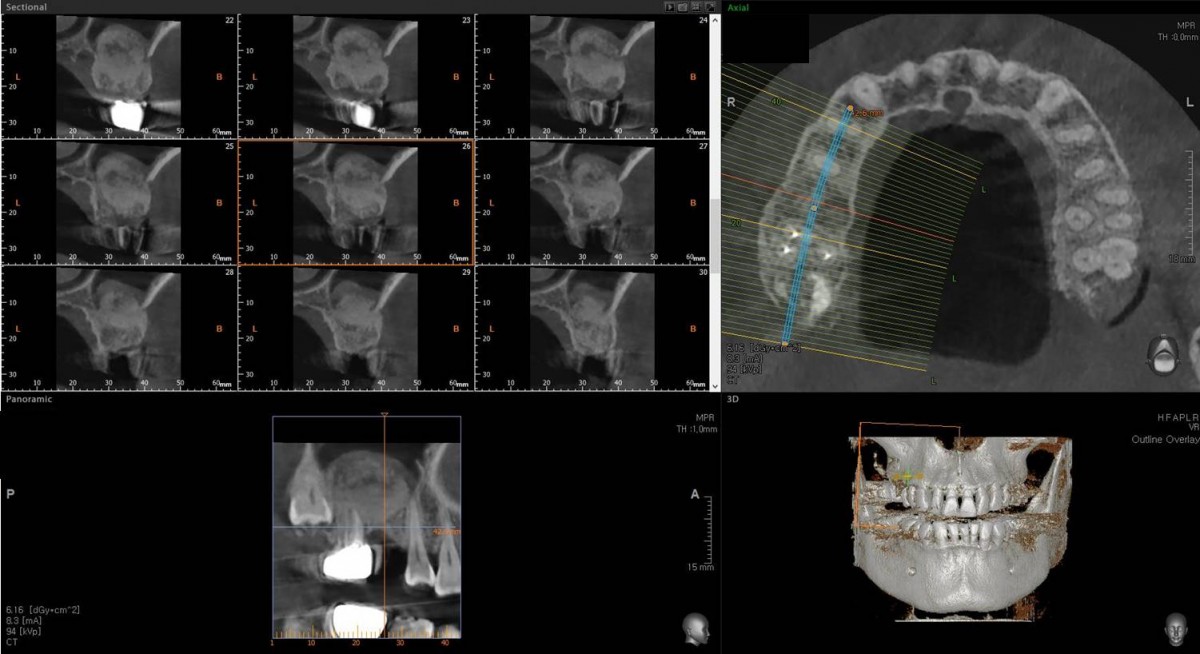 ▲CBCT after 1month of maxillary sinus graft
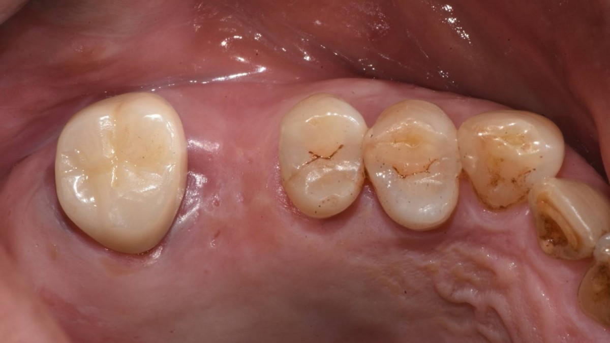 ▲4 months after sinus graft. Intra-oral view on the day of implant surgery. It was scheduled that the 2nd molar extraction and 2 implants would be placed in the 1st and 2nd molar zone.
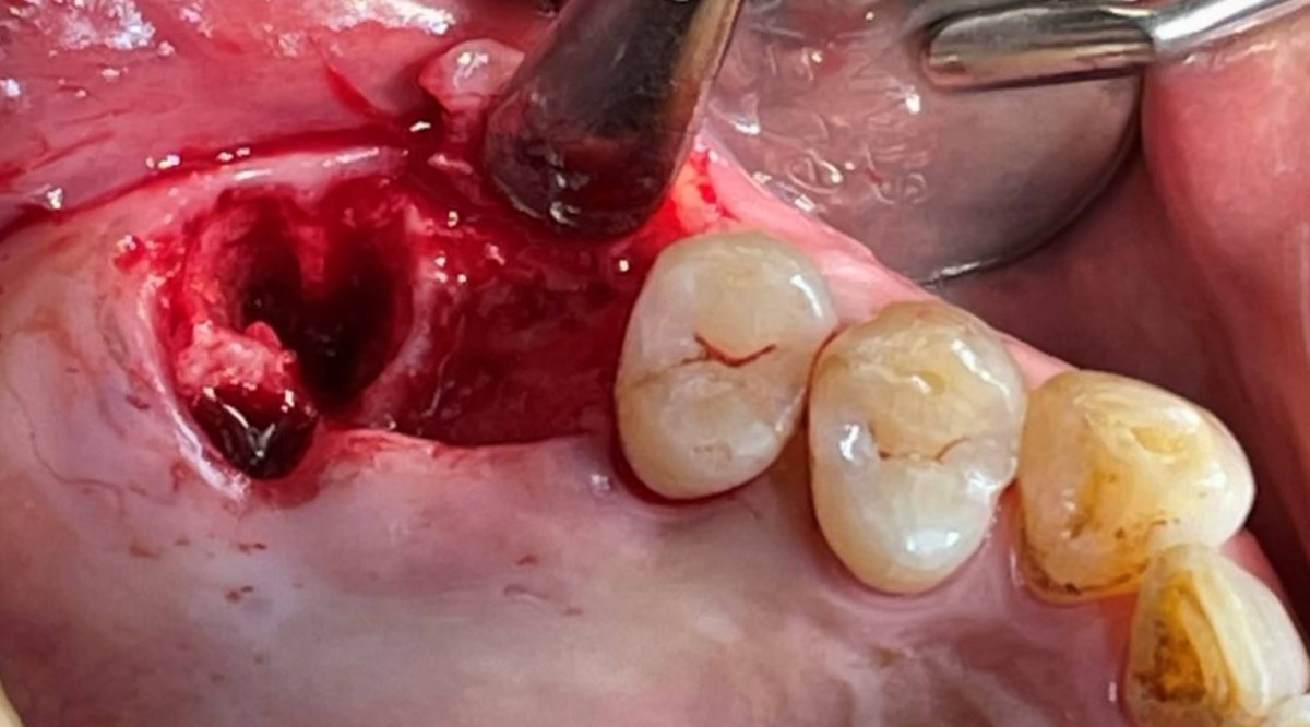 ▲The 2nd molar extraction
 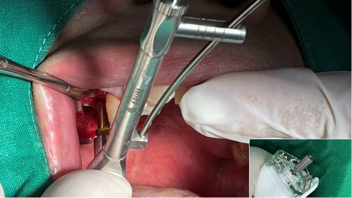 ▲Insertion torque was 35Ncm at the 1st molar zone. Arum NB1, 5*10
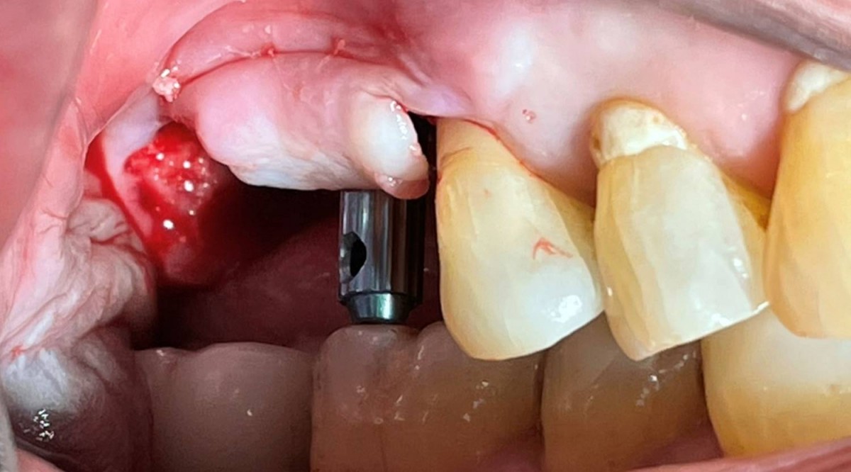 ▲Lateral view. Check the 3-dimensional position and occlusal relationship with the Arum direction pin.
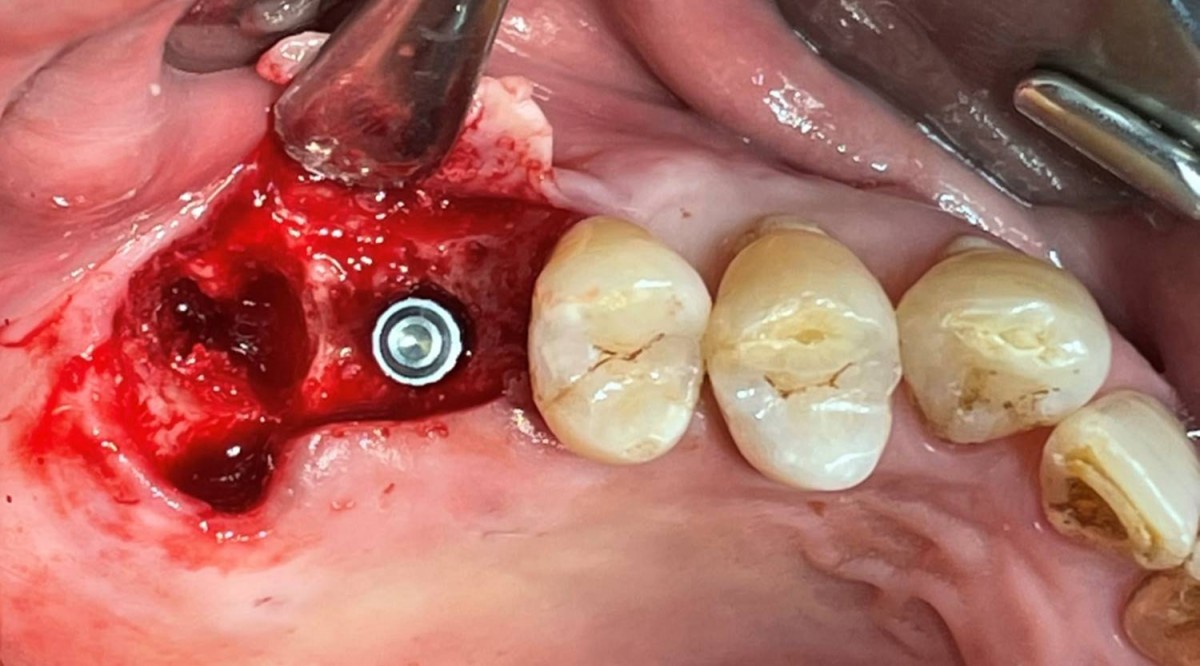 ▲Occlusal view. Check the 3-dimensional position and occlusal relationship with the Arum direction pin.
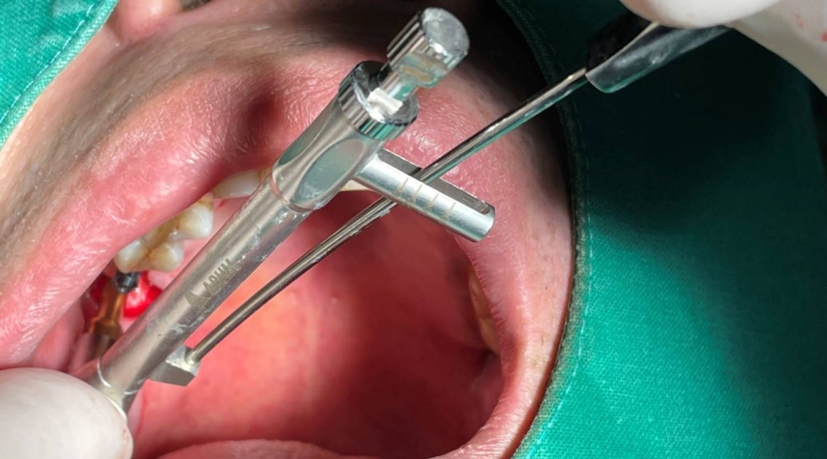 ▲Insertion torque at the 2nd molar zone was 20Ncm. Arum NB1, 5*10
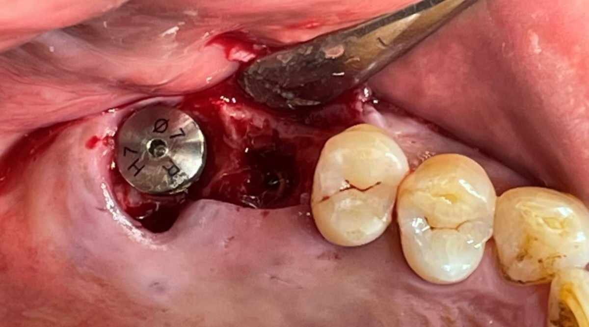 ▲A healing abutment was connected to the immediately placed implant
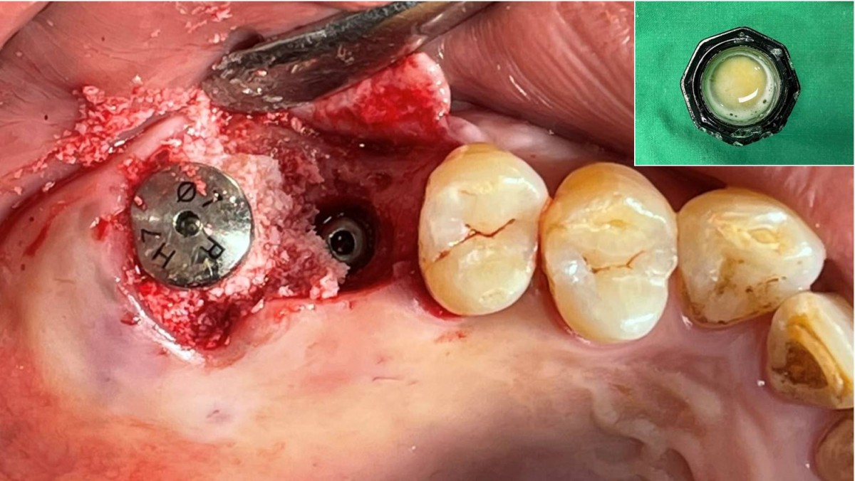 ▲GBR was performed at the immediately placed area. Xenogrtaft+CGF
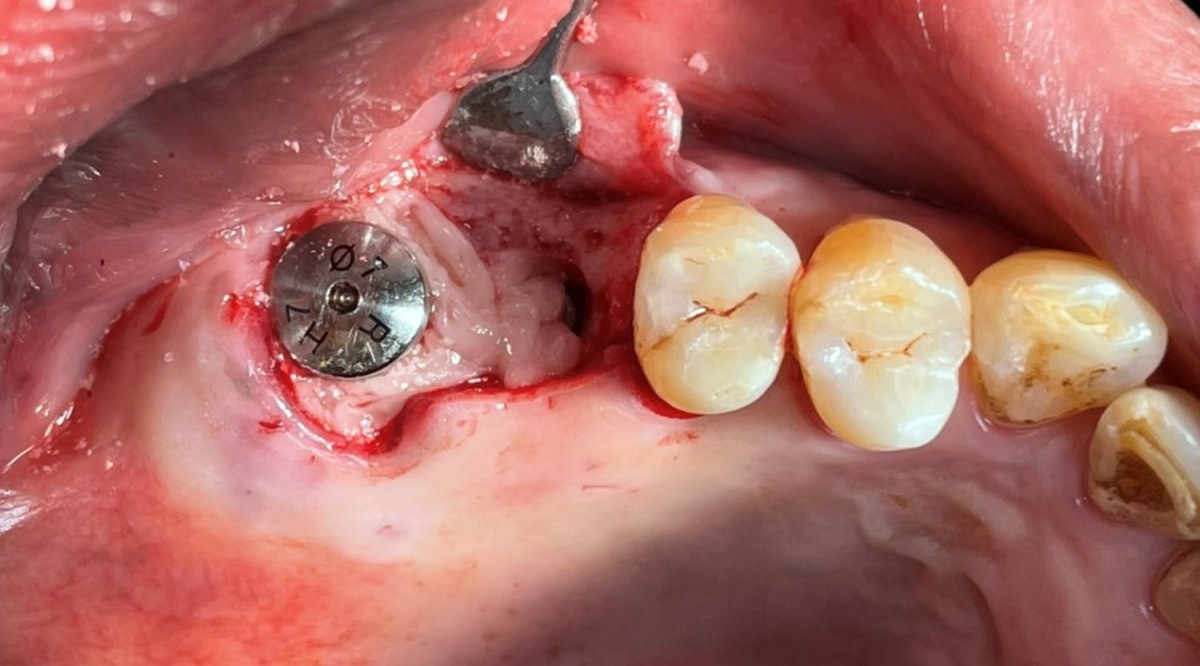 ▲CGF was placed on the grafted area
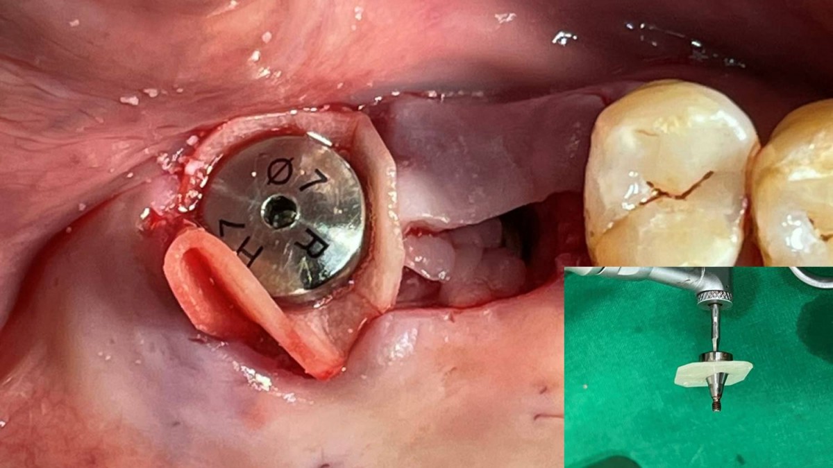 ▲A membrane-engaged healing abutment was re-screwed into the fixture after unscrewing it.
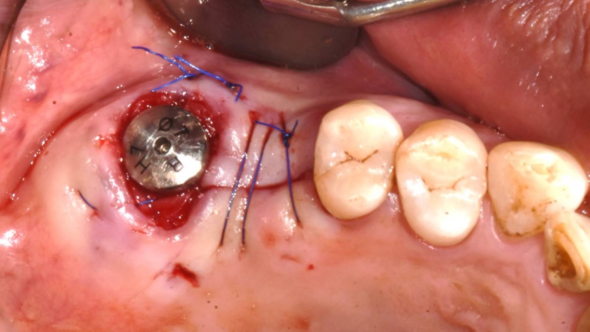 ▲The flap was closed.
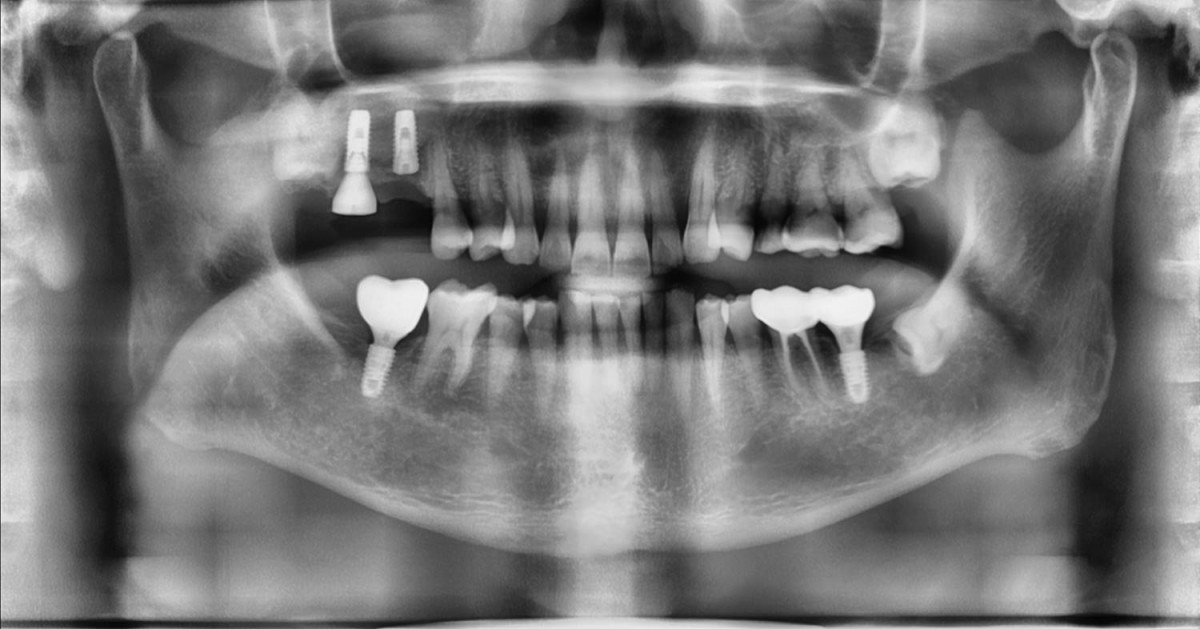 ▲ A panoramic radiograph after 2 implants were placed in the right maxilla.
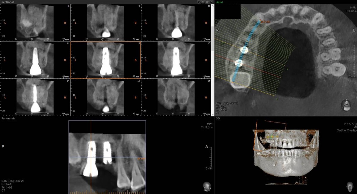 ▲CBCT scan image focused on the 2nd molar zone.
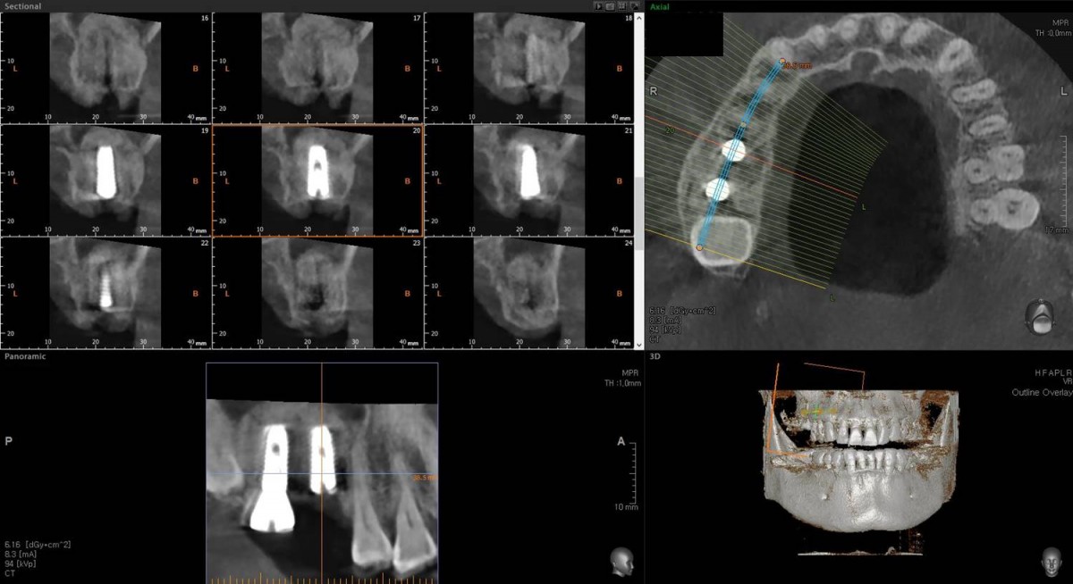 ▲CBCT scan image focused on the 1st molar zone.
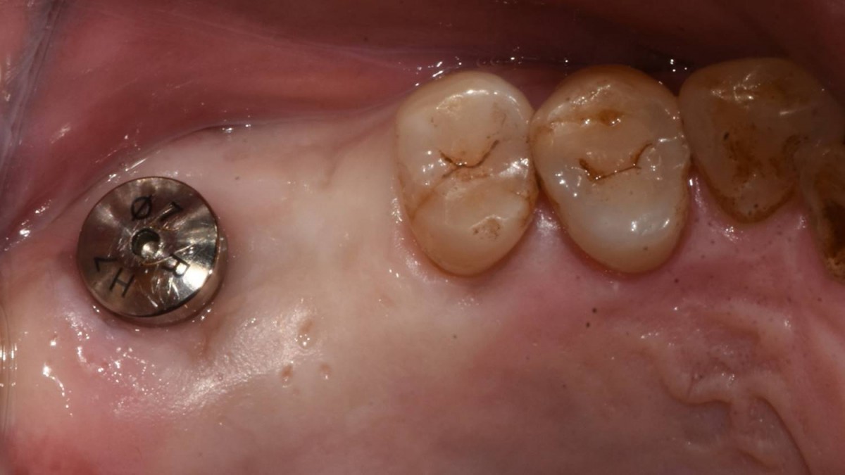 ▲4 months after implant placement. Intraoral view on the day of implant uncovery(2nd surgery).
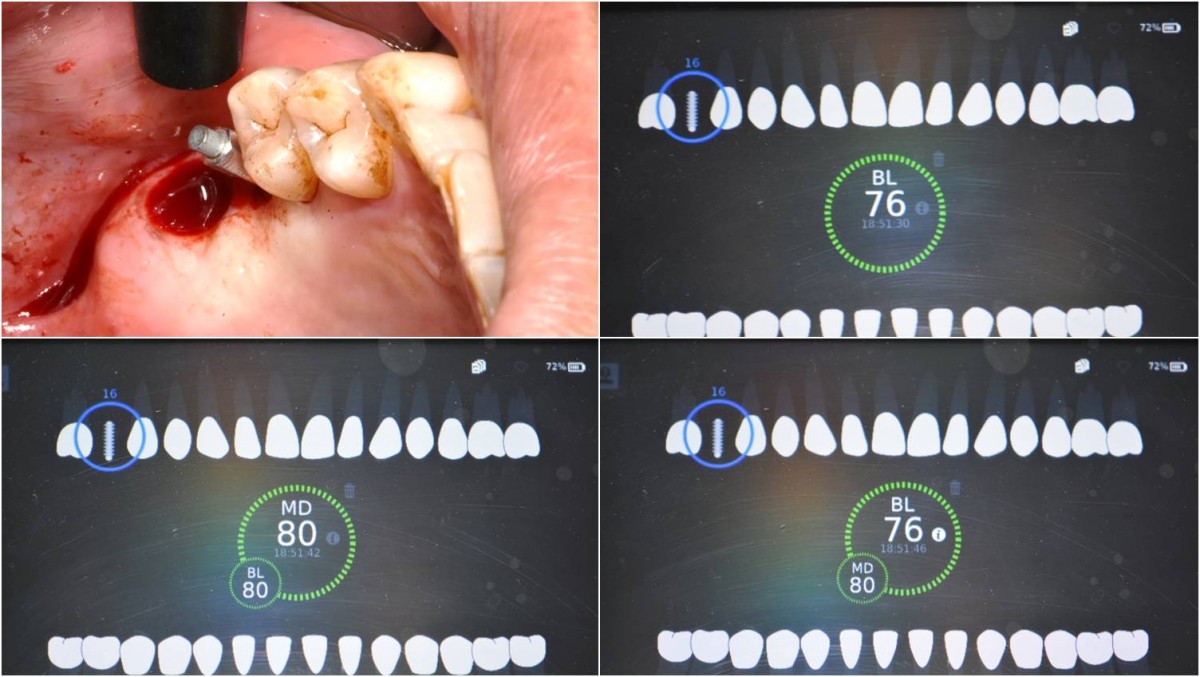 ▲4 months after implant placement. Implant uncovery and ISQ reading was performed. ISQ values at the 1st molar area.
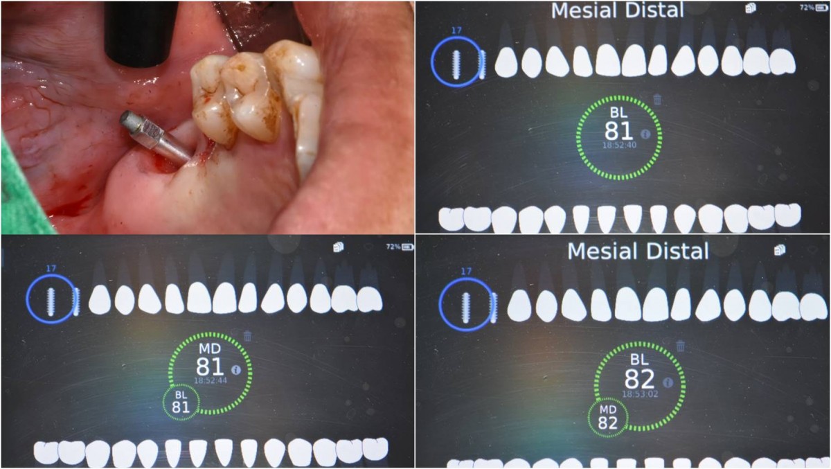 ▲ISQ values at the 2nd molar area.
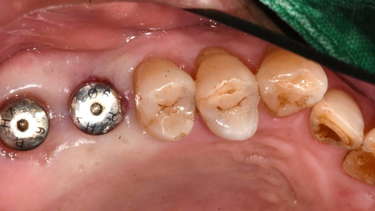 ▲After the ISQ reading, the healing abutment was engaged to the fixture.
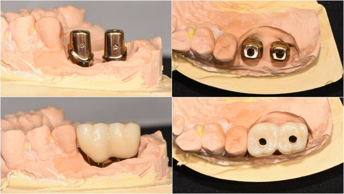 ▲When viewed from the side, the embrasure area was not opened enough. That should be re-contoured.
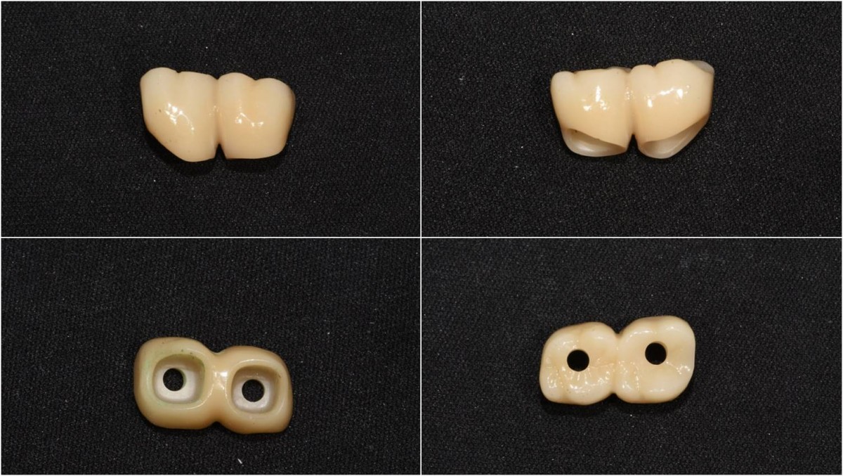 ▲The embrasure area was re-contoured.
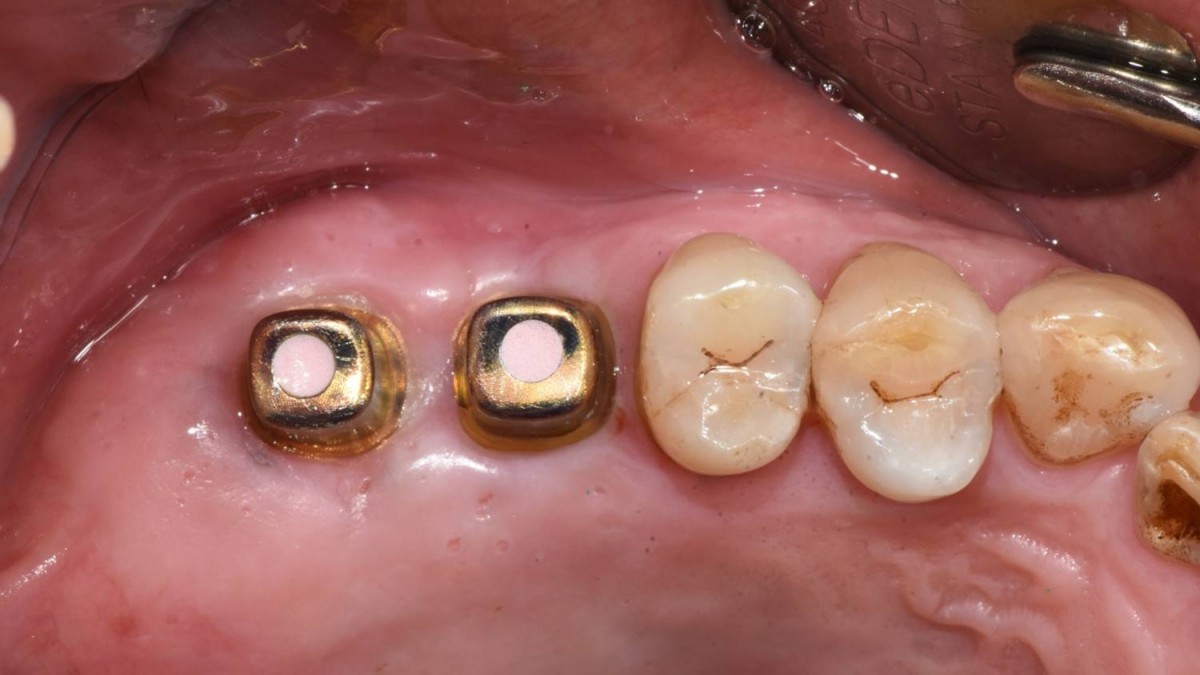 ▲Abutments were connected to fixtures
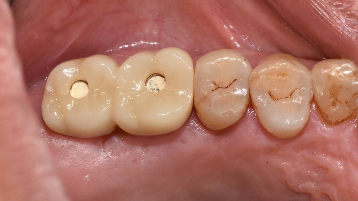 ▲Seating trial
 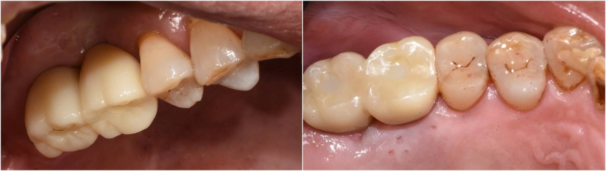 ▲ Intra-oral view after delivery (permanent cementation and access hole filling with composite resin).
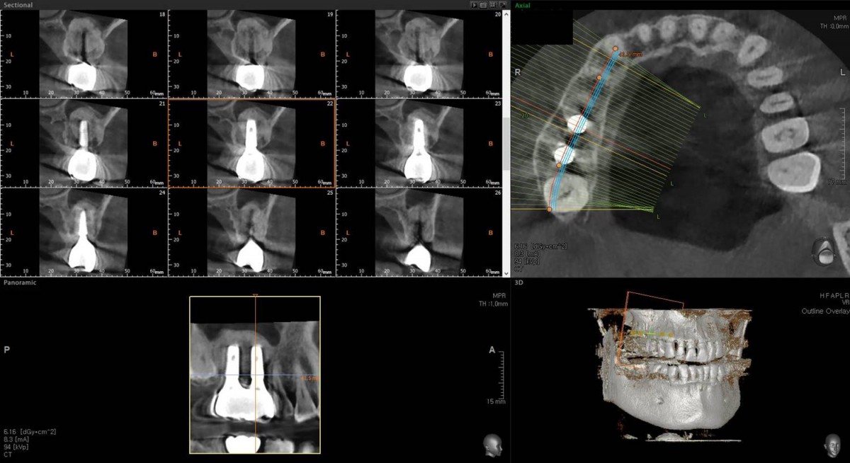 ▲CBCT scan focused on 1st molar area after crown cementation
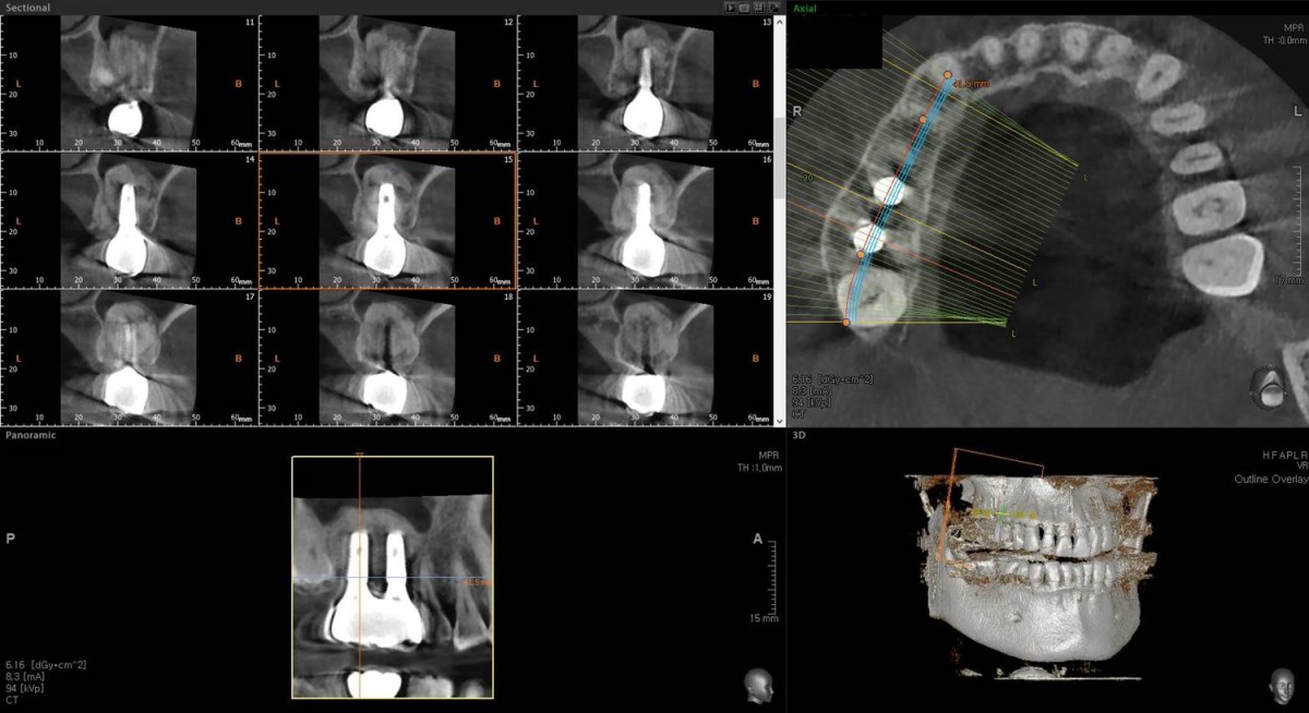 ▲CBCT scan focused on 2nd molar area after crown cementation.
0
- PrevSingle PFM crown in the esthetic regionJan, 20, 2023
- NextIn the anterior maxilla, implant-supported fixed partial denture. Jan, 20, 2023
There are no registered comment.